Removal of an intraperitoneal foreign body using a single port laparoscopic procedure
- PMID: 21902989
- PMCID: PMC3148885
- DOI: 10.4293/108680811X13071180407113
Removal of an intraperitoneal foreign body using a single port laparoscopic procedure
Erratum in
- JSLS. 2012 Jan-Mar;16(1):189
Abstract
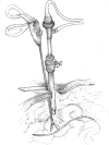
Background and objectives: To remove a foreign body from the peritoneal cavity in laparoscopic surgery, 2 or 3 ports are usually used. We have recently performed such a removal using a single 10-mm transumbilical port, a 0-degree laparoscope, a Farabeuf retractor, and a laparoscopic grasping forceps.
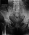
Methods: Two patients with ventriculoperitoneal shunt catheter (V-P shunt) were admitted to our unit during the last year. They previously had a shunt catheter implanted for hydrocephalus of unknown cause. The complete migration of the ventriculoperitoneal shunt catheter into the peritoneal cavity was observed in these patients 12 and 7 years after the implantation. The laparoscopic removal of the migrated catheter was decided on. Its presence and location were confirmed by the use of a 0-degree laparoscope, through a 10-mm trocar port. The catheter was held and pulled out using a grasping forceps that was pushed in just beside the trocar port.
Conclusion: The laparoscopic approach enables safe removal of a foreign body in the peritoneal cavity. The procedure can be performed using a single port.
Figures


References
-
- Esposito JM. Removal of polyethylene catheters under laparoscopic supervision. J Reprod Med. 1975;14:174. - PubMed
-
- Witmore RJ, Lehman GA. Laparoscopic removal of intraperitoneal catheter. Gastrointest Endosc. 1979;25:75–76 - PubMed
-
- Kurita N, Shimada M, Nakao T, et al. Laparoscopic removal of a foreign body in the pelvic cavity through one port using a flexible cholangioscope. Dig Surg. 2009;26:205–208 - PubMed
-
- Cope C, Kramer MS. Laparoscopic removal of dialysis catheter. Ann Intern Med. 1974;81:121. - PubMed
-
- Smith DC. Removal of an ectopic IUD through the laparoscope. Am J Obstet Gynecol. 1969;105:285–286
Publication types
MeSH terms
LinkOut - more resources
Full Text Sources
