Pre-pubertal presentation of peritoneal inclusion cyst associated with congenital lower extremity venous valve agenesis
- PMID: 21902991
- PMCID: PMC3148887
- DOI: 10.4293/108680811X13071180406835
Pre-pubertal presentation of peritoneal inclusion cyst associated with congenital lower extremity venous valve agenesis
Abstract
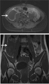
Peritoneal inclusion cysts are uncommon lesions that usually occur in the pelvis of reproductive-age females. The case of a 7-year-old girl with an inflamed peritoneal inclusion cyst with unusual right paracolic localization and congenital lower extremity superficial and deep venous valve agenesis is presented. Inflammation of the peritoneal inclusion cyst was responsible for the signs of acute abdomen and subsequent presentation at our center. The cystic structure was initially diagnosed using ultrasonography, and its complete extent (8cm x 6.5cm x 4cm) was evident after magnetic resonance imaging. The minimal access approach was opted for to resect the entire cyst from the lateral border of the ascending colon. Afterwards, the cyst was punctured to reduce its size and to retrieve the cyst wall using an endoscopic specimen retrieval bag. Minimal access surgery precautions in this patient with congenital lower extremity venous valve agenesis are discussed.
Figures


References
-
- Ross MJ, Welch WR, Scully RE. Multilocular peritoneal inclusion cysts (so-called cystic mesotheliomas). Cancer. 1989;64:1336–1346 - PubMed
-
- Sawh RN, Malpica A, Deavers MT, Liu J, Silva EG. Benign cystic mesothelioma of the peritoneum: a clinicopathologic study of 17 cases and immunohistochemical analysis of estrogen and progesterone receptor status. Hum Pathol. 2003;34:369–374 - PubMed
-
- Amesse LS, Gibbs P, Hardy J, Jones KR, Pfaff-Amesse T. Peritoneal inclusion cysts in adolescent females: a clinicopathological characterization of four cases. J Pediatr Adolesc Gynecol. 2009;22:41–48 - PubMed
-
- Urbanczyk K, Skotniczny K, Kucinski J, Friediger J. Mesothelial inclusion cysts (so-called benign cystic mesothelioma): a clinicopathological analysis of six cases. Pol J Pathology. 2005;56:81–87 - PubMed
-
- Advincula AP, Hernandez JC. Acute urinary retention caused by a large peritoneal inclusion cyst: a case report. J Reprod Med. 2006;51:202–204 - PubMed
Publication types
MeSH terms
LinkOut - more resources
Full Text Sources
Medical
