

Postpartum ovarian vein thrombosis
- PMID: 21902992
- PMCID: PMC3148888
- DOI: 10.4293/108680811X13071180406673
Postpartum ovarian vein thrombosis
Abstract
Background: Ovarian vein thrombosis (OVT) is a rare but potentially serious postpartum complication, which occurs in 0.05% to 0.18% of pregnancies and is diagnosed on the right side in 80% to 90% of the cases.
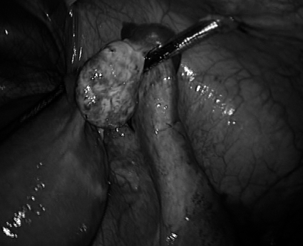
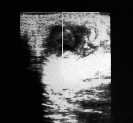
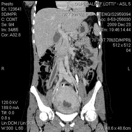
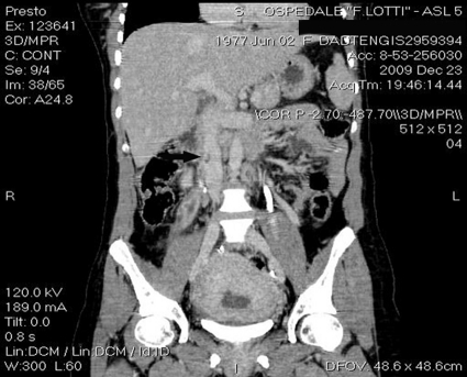
Case report: A 32-year-old woman presented at 15 days postpartum to our emergency department with severe abdominal pain, fever, and abdominal distension. Abdominal examination revealed right lower quadrant pain with rebound tenderness. The plain abdominal radiography evidenced a diffuse fecal stasis; abdominal ultrasound showed the presence of free fluid in the Douglas' pouch and between small bowel loops. Diagnosis of acute appendicitis was made. The patient immediately underwent explorative laparoscopy; at surgery, a woody tumoration consistent with right ovarian vein thrombosis was found. Laparoscopic ultrasound confirmed the diagnosis. Anticoagulation therapy and antibiotics were instituted. CT-scan confirmed the presence of thrombosis up to the vena cava. The patient was discharged on postoperative day 4. At 1-month follow-up, she remained stable and symptom free.
Discussion: Even though postpartum ovarian vein thrombosis is rare, recognition and treatment is needed to institute adequate therapy and avoid potential serious sequelae. The diagnosis can be established by ultrasound, CT scan, and MRI examinations, although, as in the case described, the limitation of ultrasound includes obscuration of the gonadic vein by overlying bowel gas.
Conclusion: OVT should be considered in any woman in the postpartum period with lower abdominal pain, fever, and leucocytosis.
Figures
References
-
- Ortin X, Ugarriza A, Espax RM, et al. Postpartum ovarian vein thrombosis. Thromb Haemost. 2005;93:1004–1005 - PubMed
-
- Dunnihoo DR, Gallaspy JW, Wise RB, Otterson WN. Post-partum ovarian vein thrombophlebitis: a review. Obstet Gynecol Surv. 1991;46:415–427 - PubMed
-
- Sinha D, Yasmin H, Samra JS. Postpartum inferior vena cava and ovarian vein thrombosis – a case report and literature review. J Obstet Gynaecol. 2005;25:312–313 - PubMed
-
- Baran GW, Frisch KM. Duplex Doppler evaluation of puerperal ovarian vein thrombosis. Am J Roentgenol. 1987;149:321–322 - PubMed
-
- Calderwood CJ, Jamienson R, Greer IA. Gestational related changes in the deep venous system of the lower limb on light reflection rheography in pregnancy and the puerperium. Clin Radiol. 2007;62:1174–1179 - PubMed
Publication types
MeSH terms
LinkOut - more resources
Full Text Sources
Medical