Review
. 2011 Sep 8;71(5):772-81.
doi: 10.1016/j.neuron.2011.07.024.

Dendritic spines and distributed circuits

Affiliations
Review

Dendritic spines and distributed circuits

Rafael Yuste. Neuron. .

Abstract

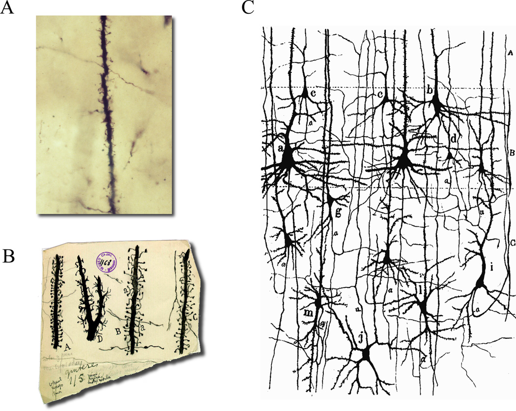
Dendritic spines receive most excitatory connections in pyramidal cells and many other principal neurons. But why do neurons use spines, when they could accommodate excitatory contacts directly on their dendritic shafts? One suggestion is that spines serve to connect with passing axons, thus increasing the connectivity of the dendrites. Another hypothesis is that spines are biochemical compartments that enable input-specific synaptic plasticity. A third possibility is that spines have an electrical role, filtering synaptic potentials and electrically isolating inputs from each other. In this review, I argue that, when viewed from the perspective of the circuit function, these three functions dovetail with one another to achieve a single overarching goal: to implement a distributed circuit with widespread connectivity. Spines would endow these circuits with nonsaturating, linear integration and input-specific learning rules, which would enable them to function as neural networks, with emergent encoding and processing of information.


Figures

Figure 1
Figure 1. Golgi stains reveal spines and straight axon
(A) Photomicrograph of an original Golgi preparation from Cajal. The image shows a segment of a dendrite from pyramidal neuron with abundant spines. In the background there are some stained axons crossing transversally. Note how the axonal trajectories are straight. (B) Cajal drawings of different types of spines. Note how spines protrude to cover the neighboring volume. Some axons are also drawn, with straight trajectories. (C) Cajal’s drawing of cellular elements of cerebral cortex. Note how axons have straight, vertical trajectories and basal dendrites are well positioned to intercept them. Reproduced with permission from “Herederos de Santiago Ramón y Cajal.”.
Figure 2
Figure 2. Helical distribution of spines
Regular features along the dendritic shaft of Purkinje cells from electric fish. (A) Spine necks forming regular linear arrays over the shaft surface, revealed in confocal sections. (B) Periodic linear arrays of spines (e.g., circles) in a dendrite. Scale bars = 1 um. (C) Diffraction pattern of B, showing two pairs of peaks arranged with mirror vertical symmetry; the distance of these peaks from the equator indicates that the periodicities repeat every 1.25 um. (D) Filtered images revealing the paths traced by lines of spines on the near sides of the dendrite, made by including only terms associated with the N pair of peaks within the masks. Reprinted with permission from O'Brien and Unwin, (2006). Organization of spines on the dendrites of Purkinje cells. Proc Natl Acad Sci U S A 103, 1575-1580. Copyright (2006) National Academy of Sciences, U.S.A. (E). Schematic rendering of a helical pattern of spines along a dendrite.
Figure 3
Figure 3. Distributed circuit model
Excitatory neurons are connected in a distributed topology, by which each cell contacts many other neurons, but makes few (or one) contact with each of them and postsynaptic cells receive inputs from many presynaptic neurons. Active neurons and inputs are black, silent cells white. Active inputs are integrated linearly and those neurons whose arithmetic input sum reaches threshold (three simultaneous inputs in this case), fire an action potential (cells 1 and 5). Meanwhile, neurons that receive a smaller number of active inputs (neurons 2, 3 and 4), fail to do so.
Figure 4
Figure 4. Input integration in vivo
Distribution of activated dendritic inputs in pyramidal cells from mouse visual cortex. Red dots indicate hotspots of local dendritic calcium signaling, evoked by drifting gratings of different orientations, superimposed on the Z-projection of the reconstructed dendritic tree. Red dashed lines point to the polar plot obtained for the corresponding local calcium signals. The frame (grey dashed line) indicates the area of imaging. Note the salt-and-pepper distribution of the orientation-tuned hot spots. The neuron was tuned for the vertical orientation, the orientation that is more represented in this sample. Reprinted by permission from Macmillan Publishers Ltd: [Nature] (Jia et al., Dendritic organization of sensory input to cortical neurons in vivo), copyright (2010).
Figure 5
Figure 5. Disordered functional circuit organization in visual cortex
Functional maps of selective responses in rat visual cortex. a, In vivo images of cortical cells stained with a calcium indicator, OGB-1 AM. The top panel shows a volume of stained cells; the bottom panel is a cell-based orientation map in which hue is determined by the best orientation overlaid with the anatomical image. Visually responsive cells are colored according to their preferred orientation (green=vertical; red=horizontal; blue and yellow=oblique). Note the apparently random spatial arrangement of orientation specificity. Scale bar, 100 um. Reprinted by permission from Macmillan Publishers Ltd: [Nature] (Ohki et al., Functional imaging with cellular resolution reveals precise micro-architecture in visual cortex), copyright (2005).

References

    1. Abeles M. Corticonics. Cambridge, England: Cambrdige University Press; 1991.
    1. Araya R, Eisenthal KB, Yuste R. Dendritic spines linearize the summation of excitatory potentials. Proc Natl Acad Sci USA. 2006a;103:18779–18804. - PMC - PubMed
    1. Araya R, Jiang J, Eisenthal KB, Yuste R. The spine neck filters membrane potentials. Proc Natl Acad Sci USA. 2006b;103:17961–17966. - PMC - PubMed
    1. Araya R, Nikolenko V, Eisenthal KB, Yuste R. Sodium channels amplify spine potentials. Proceedings of the National Academy of Sciences of the United States of America. 2007;104:12347–12352. - PMC - PubMed
    1. Arellano JI, Benavides-Piccione R, DeFelipe J, Yuste R. Ultrastructure of dendritic spines: correlation between synaptic and spine morphologies Frontiers. Neuroscience. 2007a;1:131–143. - PMC - PubMed

Publication types

LinkOut - more resources