. 2011 Sep 8;71(5):799-811.
doi: 10.1016/j.neuron.2011.07.022.

Development of a method for the purification and culture of rodent astrocytes

Affiliations

Development of a method for the purification and culture of rodent astrocytes

Lynette C Foo et al. Neuron. .

Abstract

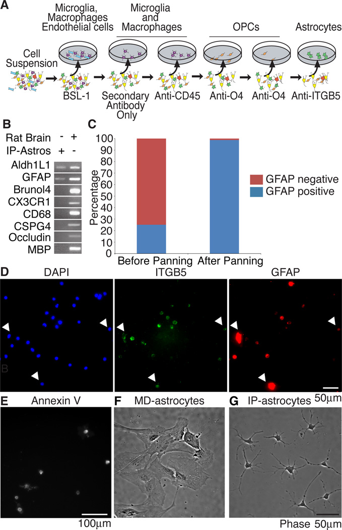
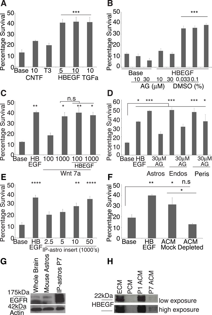
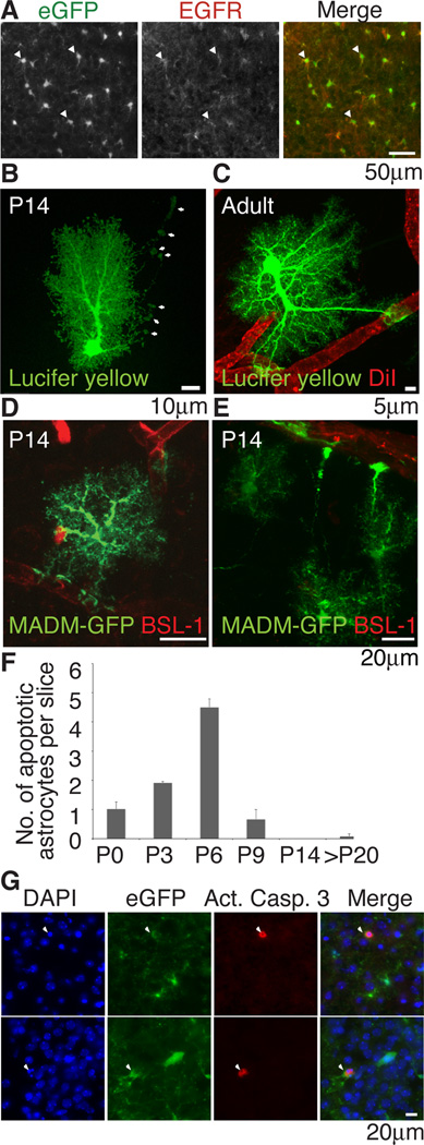
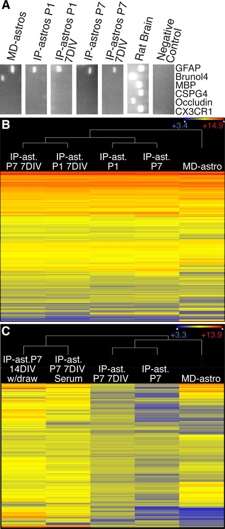
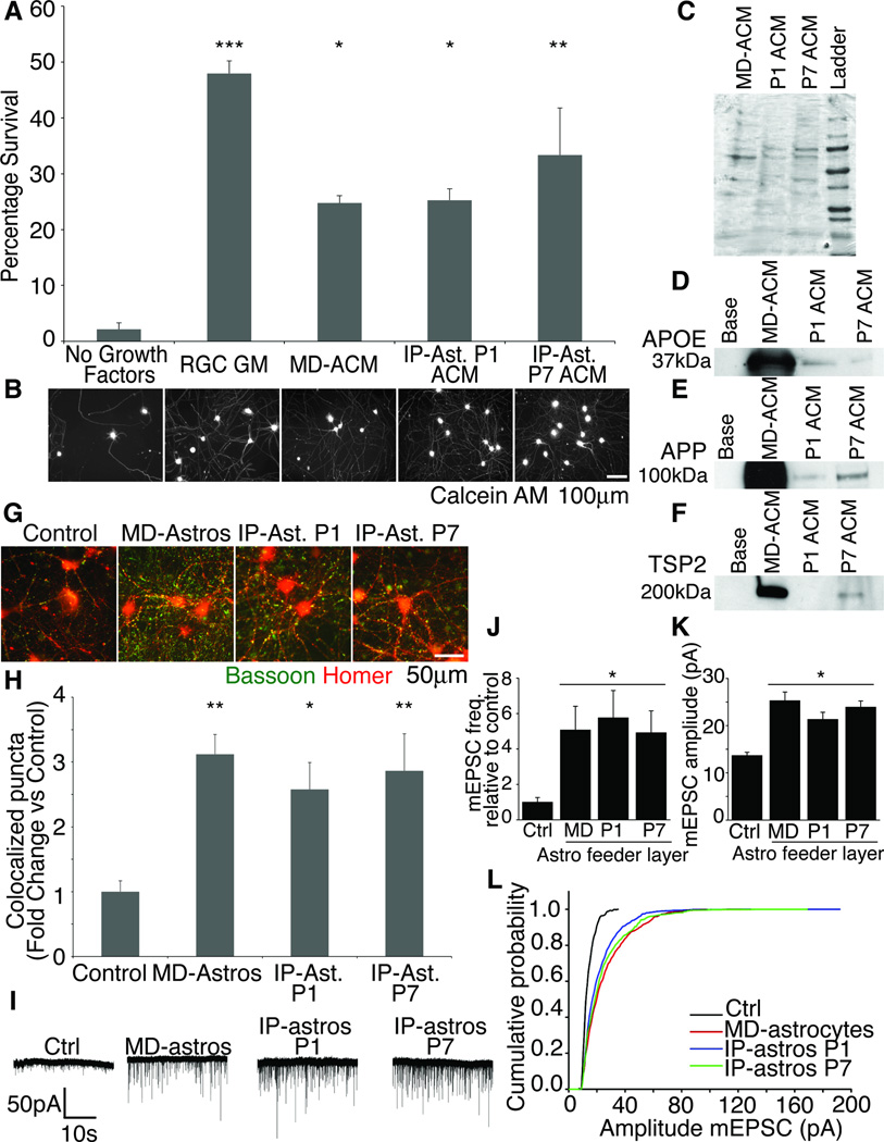
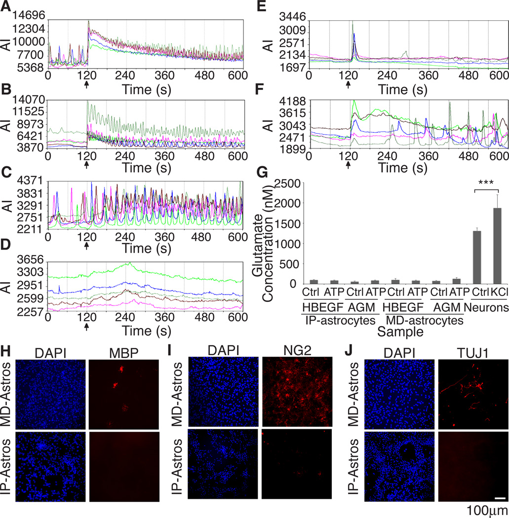
The inability to purify and culture astrocytes has long hindered studies of their function. Whereas astrocyte progenitor cells can be cultured from neonatal brain, culture of mature astrocytes from postnatal brain has not been possible. Here, we report a new method to prospectively purify astrocytes by immunopanning. These astrocytes undergo apoptosis in culture, but vascular cells and HBEGF promote their survival in serum-free culture. We found that some developing astrocytes normally undergo apoptosis in vivo and that the vast majority of astrocytes contact blood vessels, suggesting that astrocytes are matched to blood vessels by competing for vascular-derived trophic factors such as HBEGF. Compared to traditional astrocyte cultures, the gene profiles of the cultured purified postnatal astrocytes much more closely resemble those of in vivo astrocytes. Although these astrocytes strongly promote synapse formation and function, they do not secrete glutamate in response to stimulation.


Figures

Figure 1
Figure 1. Establishment of an immunopanning protocol for rat astrocytes
(A) Cortical suspensions were passed over successive panning plates to remove endothelial cells and microglia (BSL-1), microglia and macrophages (secondary only plate), microglia (CD45), oligodendrocyte precursor cells (O4) and finally a positive panning plate for astrocytes (ITGB5). (B) Purity of IP-astrocytes validated by RT-PCR and (C) immunostaining with GFAP. Before immunopanning, the whole cell brain suspension contained 25.1% GFAP+ cells, after immunopanning, the isolated cells were 98.7% GFAP+. (D) ITGB5 was present on all astrocytes. All cells in cortical cell suspensions that were GFAP+ were also ITGB5+ (arrowheads). (E) Astrocytes are positive for the apoptotic marker Annexin V. (F) MD-astrocytes had flat and fibroblast-like morphologies at 14DIV (G) IP-astrocytes cultured with HBEGF for 14DIV were healthy and extended multiple processes.
Figure 2
Figure 2. P7 Astrocytes require trophic factors for survival or die by apoptosis
(A) Addition of 5 or 10 ng/ml of HBEGF or 10ng/ml TGFα significantly (p<0.001) promoted IP-astrocyte survival. Neither 10ng/ml CNTF nor T3 significantly increased survival of IP-astrocytes. (B) AG1478 (AG), did not affect basal survival of astrocytes, but at 10µM and 30µM, significantly inhibited the effect of 5ng/ml HBEGF (HBEGF) not observed in control conditions with DMSO (p<0.001). (C) 1µg/ml of Wnt7a is a trophic factor for astrocytes (**p<0.01), but the effect was not significantly additive with 5ng/ml HBEGF. Values on x-axis are in ng/ml (D) Feeder layers of astrocytes, endothelial cells and pericytes produced soluble trophic factors that significantly promoted the survival of astrocytes (***p<0.001). 30µM AG1478 (AG) negated the survival-promoting effect of astrocytes and partially of endothelial cells, but not pericytes (*p<0.05). (E) IP-astrocytes P7 grown in inserts at densities higher than 10,000 cells/insert kept IP-astrocytes P7 alive. (F) Mock depletion of ACM with goat anti-Gγ13 IgG did not negate the survival-promoting effect of ACM. Depletion of ACM with goat anti-HBEGF IgG negated the survival-promoting effect of ACM. (G) Western blot analysis revealed that both mouse astrocytes and rat IP-astrocytes P7 expressed EGFR. (H) ECM and P1 ACM contained high levels of HBEGF, P7 ACM contained lower levels and none in PCM. Top blot: low exposure, bottom blot: high exposure. Also see Figure S1
Figure 3
Figure 3. Astrocytes express EGFR, contact blood vessels and die by apoptosis in vivo
(A) Immunohistochemical staining for EGFR in P6 Aldh1L1-eGFP mice showed that majority of cortical astrocytes expressed EGFR (white arrowheads=EGFR+,Aldh1L1-eGFP+). (B,C) All hippocampal astrocytes at P14 (n=23) and adult (n=22) contacted blood vessels. (B) P14 astrocytes send out long processes that contact but not ensheathed blood vessels (white arrows). (C) Full ensheathement was observed in the adult. (D,E) MADM-labeling sparsely labels astrocytes eGFP+ in the brain. Blood vessels were stained with BSL-1 and eGFP+ astrocytes of the cortex visualized with confocal microscopy. 30 of 31 astrocytes visualized contacted blood vessels (F,G) Quantification of immunohistochemical staining with activated caspase 3 (red) in Aldh1L1-eGFP (green) mice and colocalization with condensed nuclei visualized with DAPI (blue) (arrowheads) revealed that astrocyte apoptosis was observed at P6 (G) Immunostaining of apoptotic astrocytes in P6 Aldh1L1-eGFP mice.
Figure 4
Figure 4. Comparison of IP-astrocyte expression profiles with MD-astrocytes
(A) Purity was assessed by RT-PCR with major cell type markers. MD-astrocytes have neuronal contamination not observed in IP-astrocytes whether acute or cultured. (B) Heat map of 15,960 genes where gene expression exceeds 200 in at least one sample. Acutely isolated IP-astrocytes at P1 and P7 (IP-ast P1, P7), IP-astrocytes that have been cultured in HBEGF for 7 days (IP-ast P1, P7 7DIV) and MD-astrocytes gene expression was normalized and plotted on a log2 color scale. Cooler colors represent low expression and warm colors the converse. MD-astrocytes profiles are distinct from IP-astrocytes and cultured IP-astrocytes are closer in their profiles to their acutely isolated counterparts. (C) Heat map showing expression levels of 365 serum-induced genes across samples. IP-Astrocytes P7 were treated for 7d immediately after isolation with 10% serum and either processed for RNA (IP-ast P7 7DIV Serum) or the serum withdrawn and cells cultured for an additional 7d (IP-ast P7 14DIV w/draw) and compared to IP-ast. P7 7DIV cultured in base media with HBEGF, acutely isolated IP-ast P7 and MD-astrocytes. Serum induction did not cause IP-astrocytes to exhibit a profile like MD-Astrocytes and serum withdrawal did not cause reversion of the serum-induced genes. Also see Tables S1–5.
Figure 5
Figure 5. IP-astrocytes in culture retained functional properties
(A,B) IP-astrocyte ACM was as capable of keeping neurons alive as MD-astrocytes was. The neurons were healthy and extended multiple processes. Majority of neurons died in the absence of trophic support. ACM derived from IP-astrocytes P1 and P7 (IP-ast P1 and P7 ACM), MD-astrocytes (MD-ACM) and a positive control of RGC growth media was used. (C) Coomassie gel of ACM used to ensure equivalent protein loading. (D) MD-astrocytes produced much higher levels of APOE (D), APP (E) and TSP2 (F), compared to P1 and P7 ACM. P1 ACM did not contain detectable levels of TSP2. (G) Synaptogenesis was quantified by assessing colocalization of presynaptic marker bassoon (green) and postsynaptic marker homer (red) with ImageJ. (H) IP-ast P1 and P7 feeder layers were as effective at inducing structural synapses as MD-astrocytes were. Without an astrocyte feeder layer, few synapses were observed (control) (**p<0.01,*p<0.05) (I) Sample traces of whole-cell patch clamp recordings from RGCs made in the presence of TTX. Few mEPSCs were observed without feeder layer of astrocytes (Ctrl). (J) Frequency and amplitude of mEPSCs recorded increased significantly with MD-astrocytes, IP-astros P1 or P7 feeder layers (p <0.05). (L) IP-astros P1 and P7 caused a shift in cumulative amplitude of mEPSCs to a similar level as MD-astrocytes.
Figure 6
Figure 6. Calcium responses to different stimuli differ between MD-astrocytes and IP-astrocytes and MD-astrocytes are contaminated with several cell types
Astrocytes do not exhibit glutamate release in response to ATP in vitro (A–F) Stimuli was added at 120s (black arrow). Graphs of calcium responses from 5 different cells. Graph axes are average intensity (AI, arbitrary units) vs time (s) (A) Both MD-astrocytes and (B) IP-astrocytes P7 responded to ATP with increased calcium oscillations. (C) MD-astrocytes responded (83.4±4.4%, n=118, p<0.0001) robustly to 50mM KCl with increased frequency of oscillations. (D) No calcium response was observed with KCl addition in IP-astrocyte cultures. (E) No response of cells due to media addition was observed in IP-astrocytes treated with 10% serum for 4 days. (F) Cultured IP-astrocytes treated with 10% serum caused a significant number of astrocytes to respond to KCl (53.3±7.4%, n=209, p<0.001). (G) Glutamate was readily released by neurons with KCl stimulation (***p<0.001). Release was not detected in IP nor MD-astrocytes treated with HBEGF or MD-astrocyte growth media (AGM,10% serum) in response to 100µM ATP. (H–J) MD-astrocyte cultures were contaminated with oligodendrocytes (MBP), OPCs and pericytes (NG2) and neurons (TUJ1) whereas minimal contamination was observed in cultures of IP-astrocytes. Also see Figure S2.

References

    1. Aguado F, Espinosa-Parrilla JF, Carmona MA, Soriano E. Neuronal Activity Regulates Correlated Network Properties of Spontaneous Calcium Transients in Astrocytes In Situ. J. Neurosci. 2002;22:9430–9444. - PMC - PubMed
    1. al-Ali SY, al-Hussain SM. An ultrastructural study of the phagocytic activity of astrocytes in adult rat brain. Journal of anatomy. 1996;188(Pt 2):257–262. - PMC - PubMed
    1. Banker GA. Trophic interactions between astroglial cells and hippocampal neurons in culture. Science. 1980;209:809–810. - PubMed
    1. Barres BA, Silverstein BE, Corey DP, Chun LL. Immunological, morphological, and electrophysiological variation among retinal ganglion cells purified by panning. Neuron. 1988;1:791–803. - PubMed
    1. Barres BA, Hart IK, Coles HS, Burne JF, Voyvodic JT, Richardson WD, Raff MC. Cell death and control of cell survival in the oligodendrocyte lineage. Cell. 1992;70:31–46. - PubMed

Publication types

MeSH terms

Associated data