Review
. 2012 Jan;133(1):40-69.
doi: 10.1016/j.pharmthera.2011.08.001. Epub 2011 Aug 26.

G protein-coupled receptor kinases: more than just kinases and not only for GPCRs

Affiliations
Review

G protein-coupled receptor kinases: more than just kinases and not only for GPCRs

Eugenia V Gurevich et al. Pharmacol Ther. 2012 Jan.

Abstract

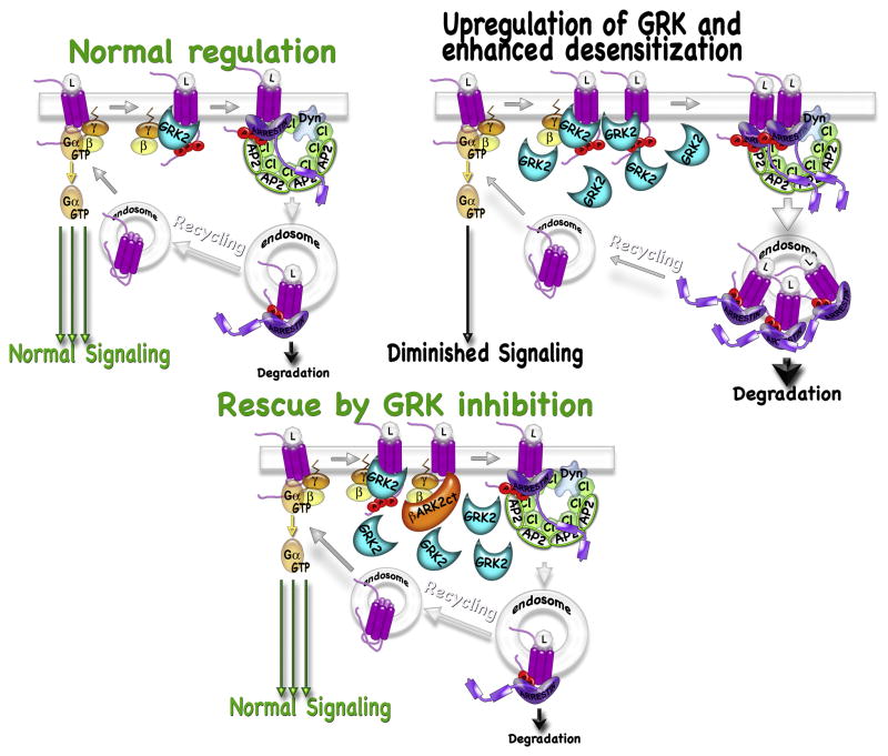
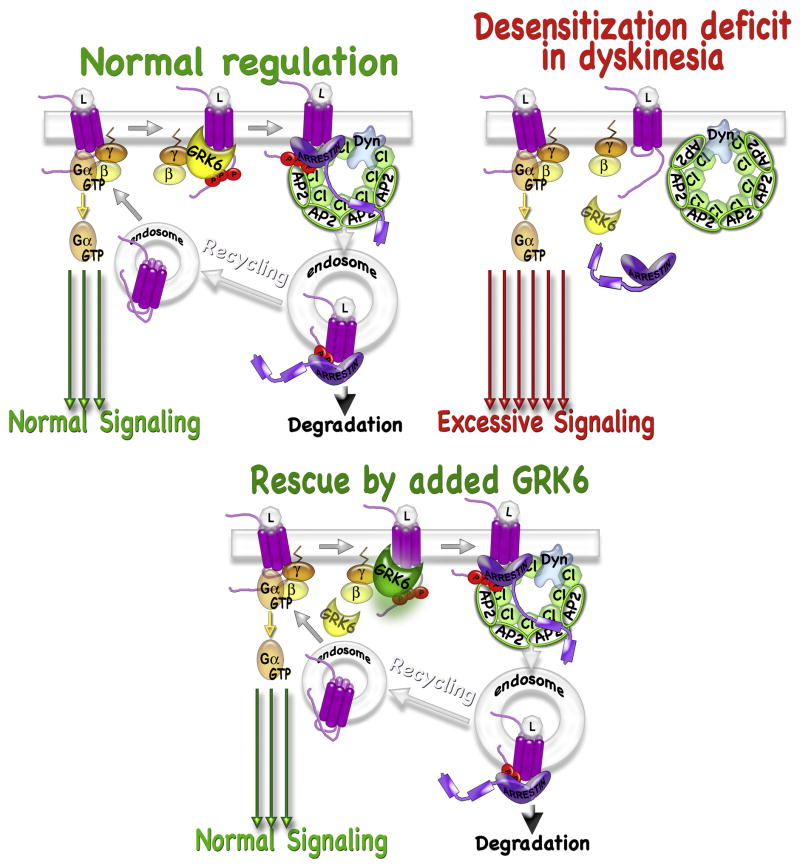
G protein-coupled receptor (GPCR) kinases (GRKs) are best known for their role in homologous desensitization of GPCRs. GRKs phosphorylate activated receptors and promote high affinity binding of arrestins, which precludes G protein coupling. GRKs have a multidomain structure, with the kinase domain inserted into a loop of a regulator of G protein signaling homology domain. Unlike many other kinases, GRKs do not need to be phosphorylated in their activation loop to achieve an activated state. Instead, they are directly activated by docking with active GPCRs. In this manner they are able to selectively phosphorylate Ser/Thr residues on only the activated form of the receptor, unlike related kinases such as protein kinase A. GRKs also phosphorylate a variety of non-GPCR substrates and regulate several signaling pathways via direct interactions with other proteins in a phosphorylation-independent manner. Multiple GRK subtypes are present in virtually every animal cell, with the highest expression levels found in neurons, with their extensive and complex signal regulation. Insufficient or excessive GRK activity was implicated in a variety of human disorders, ranging from heart failure to depression to Parkinson's disease. As key regulators of GPCR-dependent and -independent signaling pathways, GRKs are emerging drug targets and promising molecular tools for therapy. Targeted modulation of expression and/or of activity of several GRK isoforms for therapeutic purposes was recently validated in cardiac disorders and Parkinson's disease.


Conflict of interest statement

Conflict of Interest Statement

The authors declare that there are no conflicts of interests.

Figures

Figure 1
Figure 1. Domain structure of GRKs
Number above the structures indicate amino acid residue numbers of human GRKs based on (Lodowski et al., 2006). All GRKs have a short N-terminal region (green), which is implicated in GPCR binding, followed by RGS homology (RH) domain (magenta). This N-terminal region is unique to the GRK family of kinases. The RH domain is interrupted by the catalytic domain shared by all kinases (dark yellow). These elements are shared by the GRK2/3 and GRK4/5/6 subfamilies. The defining feature of the GRK2/3 subfamily is a C-terminal pleckstrin homology (PH) domain (blue) implicated in binding anionic phospholipids and Gβγ. Members of GRK4/5/6 subfamily use alternative mechanisms for membrane targeting, which include palmitoylation [palmitoylation sites are shown for GRK6A (Jiang et al., 2007)], patches of positively charged residues [amphipathic helix motifs (Thiyagarajan et al., 2004; Jiang et al., 2007) are shown as green boxes; N-terminal basic patches (Pitcher et al., 1996; Boguth et al., 2010) are shown as red boxes], and, in case of visual subtypes, prenylation (C-terminal prenylation sites in GRK1 and 7 are shown as red triangles). Residues Arg106 and Asp110 in GRK2/3, among others, are important for binding Gαq, a function unique to this subfamily. The position of the key lysine responsible for catalysis in the kinase domain is shown. Mutations K220R in GRK2 and 3, as well as K216M/K217M (Sallese et al., 2000b) in GRK4, K415 in GRK5 (Tiruppathi et al., 2000), and K215M/K216M in GRK 6(Lazari et al., 1999) yield kinase-dead GRKs. The blue box shows the position of the nuclear localization signal (NLS) in GRK5 (residues 388–395) (Johnson et al., 2004). Splice variants of GRK 4 (GRK4β, GRK4γ, and GRK4δ) are produced by in-frame deletion of exon 2 (GRK4β), exon 15 (GRK4γ), or both (GRK4δ (Premont et al., 1996; Sallese et al., 1997; Premont et al., 1999) (gene structure is shown under GRK4 protein; the exons not used in all splice variants are shown in blue) GRK6 splice variants are produced by a frame shift in the C-terminus resulting in a completely different C-terminal sequence in GRK6B as compared to GRK6A and in premature transcription termination in GRK6C (Premont et al., 1999). To generate GRK6A, exon 16 starts two nucleotides downstream, as compared to the longest variant GRK6B, resulting in a frame shif. An alternative upstream exon 16 encoding one amino acid before the stop codon is used to generate GRK6C (respective exons are labeled Exon 16A, Exon 16B, and Exon 16C). In the C-termini of GRK6 splice variants amphipathic helix residues are shown in blue and palmitoylated cysteine in red. Note the lack of palmitoylation sites in GRK6B or GRK6C.
Figure 2
Figure 2. Pathological consequences of enhanced GRK activity and potential therapeutic approaches
Normal regulation of receptor sensitivity involves receptor phosphorylation by a GRK followed by high affinity binding of an arrestin. Arrestin mediates receptor internalization via coated pits followed by either receptor recycling or degradation. In some pathological cases such as heart failure, an upregulation of one or more GRK isoforms can occur. Increased GRK availability leads to receptor hyper-phosphorylation, facilitated receptor desensitization, internalization, and, ultimately, excessive degradation. Similar effect might be achieved when a GRK with a polymorphism yielding higher activity is expressed. Such a pathological phenotype might be rescued by expression of a GRK inhibitor. Here the use of βARKct is shown, which competes with GRK2 and 3 for Gβγ, thus impeding GRK recruitment to active receptors. Alternatively, GRK knockdown could be used.
Figure 3
Figure 3. Pathological consequences of reduced GRK activity and potential therapeutic approaches
In some pathological cases exemplified by L-DOPA-induced dyskinesia, a reduced activity of one or more GRK isoforms seems to be the culprit resulting in supersensitivity of dopaminergic receptors to dopamine stimulation. A defect in receptor desensitization and/or trafficking is evidenced by persistence of D1 dopamine receptors at the plasma membrane, whereas in control animals the receptors are internalized upon L-DOPA administration (Guigoni et al., 2007; Ahmed et al., 2010). The deficiency in GRK function need not necessarily be due to reduced expression, but might stem from insufficient GRK availability in the face of increased demand. In case of dyskinesia, human patients with Parkinson’s disease or animals with experimental parkinsonism are treated with L-DOPA, which produces surges of dopamine in the brain putting pressure on regulatory signaling mechanisms. Overexpression of an appropriate GRK (e.g., GRK6A was successfully used for L-DOPA-induced dyskinesia (Ahmed et al., 2010)] might relieve that pressure by facilitating receptor desensitization and normal trafficking. Alternatively, positive allosteric modulators of GRKs might be developed. L-DOPA-induced dyskinesia is a good example of a condition where simply shutting down signaling by inhibiting dopamine receptors is not an option, because that would defeat the purpose of L-DOPA treatment, which is to provide dopaminergic stimulation to relieve akinesia in Parkinson’s patients. In contrast, using GRK as a target allows for judicial reduction in dopaminergic signaling that alleviates dyskinesia while preserving therapeutic activity of the drug.

References

    1. Ahmed MR, Bychkov E, Gurevich VV, Benovic JL, Gurevich EV. Altered expression and subcellular distribution of GRK subtypes in the dopamine-depleted rat basal ganglia is not normalized by l-DOPA treatment. J Neurochem. 2007;104:1622–1636. - PMC - PubMed
    1. Ahmed MR, Gurevich VV, Dalby KN, Benovic JL, Gurevich EV. Haloperidol and clozapine differentially affect the expression of arrestins, receptor kinases, and ERK activation. J Pharmacol Exp Ther. 2008 jpet.107.131987. - PMC - PubMed
    1. Ahmed MR, Berthet A, Bychkov E, Porras G, Li Q, Bioulac BH, et al. Lentiviral overexpression of GRK6 alleviates L-dopa-induced dyskinesia in experimental Parkinson’s disease. Sci Transl Med. 2010;2:28ra28. - PMC - PubMed
    1. Alloway PG, Howard L, Dolph PJ. The formation of stable rhodopsin-arrestin complexes induces apoptosis and photoreceptor cell degeneration. Neuron. 2000;28:129–138. - PubMed
    1. Ambrose C, James M, Barnes G, Lin C, Bates G, Altherr M, et al. A novel G protein-coupled receptor kinase gene cloned from 4p16.3. Hum Mol Genet. 1992;1:697–703. - PubMed

Publication types

MeSH terms

Substances