

Perinatal infection, inflammation, and retinopathy of prematurity
- PMID: 21903492
- PMCID: PMC3242877
- DOI: 10.1016/j.siny.2011.08.007
Perinatal infection, inflammation, and retinopathy of prematurity
Abstract
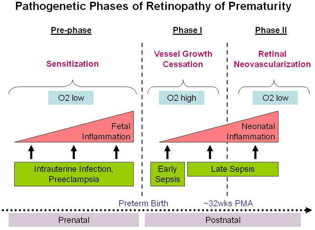
The major known risk factors for retinopathy of prematurity (ROP) are extremely low gestational age, exposure to high levels of oxygen early after birth (phase I) and relatively lower oxygen levels later (phase II). In this review, we summarize recent data suggesting that exposure to perinatal infection/inflammation is associated with an increased risk for ROP. Part of this effect might be due to direct exposure of the developing retina to circulating products of infection and/or inflammation. Another potential mechanism that deserves exploration is that inflammation and/or oxidative stress can modify the known increased risk of oxygen-associated ROP. Taken together, accumulating evidence suggests that prenatal, perinatal, and postnatal systemic inflammation contribute to a 'pre-phase', sensitizing the pre-ROP retina for subsequent insults, setting the stage for what are now called phase I and phase II of ROP pathogenesis. Strategies targeting inflammatory responses might help reduce the risk for ROP in extremely low gestational age newborns.
Copyright © 2011 Elsevier Ltd. All rights reserved.
Conflict of interest statement
None declared.
Figures
References
-
- McGregor ML, Bremer DL, Cole C, et al. Retinopathy of prematurity outcome in infants with prethreshold retinopathy of prematurity and oxygen saturation >94% in room air: the high oxygen percentage in retinopathy of prematurity study. Pediatrics. 2002;110:540–544. - PubMed
-
- Paysse EA, Lindsey JL, Coats DK, Contant CF, Jr, Steinkuller PG. Therapeutic outcomes of cryotherapy versus transpupillary diode laser photocoagulation for threshold retinopathy of prematurity. J AAPOS. 1999;3:234–240. - PubMed
-
- Gilbert C. Retinopathy of prematurity: a global perspective of the epidemics, population of babies at risk and implications for control. Early Hum Dev. 2008;84:77–82. - PubMed
-
- Chen J, Smith LE. Retinopathy of prematurity. Angiogenesis. 2007;10:133–140. - PubMed
Publication types
MeSH terms
Grants and funding
LinkOut - more resources
Full Text Sources
