. 2011 Dec;301(6):G981-91.
doi: 10.1152/ajpgi.00061.2011. Epub 2011 Sep 8.

Castration inhibits biliary proliferation induced by bile duct obstruction: novel role for the autocrine trophic effect of testosterone

Affiliations

Castration inhibits biliary proliferation induced by bile duct obstruction: novel role for the autocrine trophic effect of testosterone

Fuquan Yang et al. Am J Physiol Gastrointest Liver Physiol. 2011 Dec.

Abstract

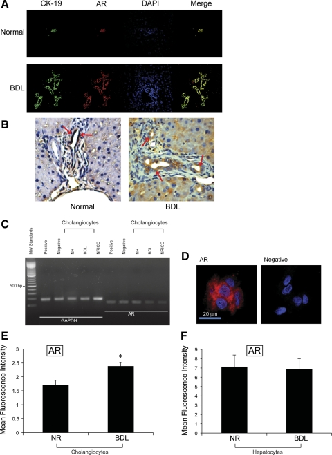
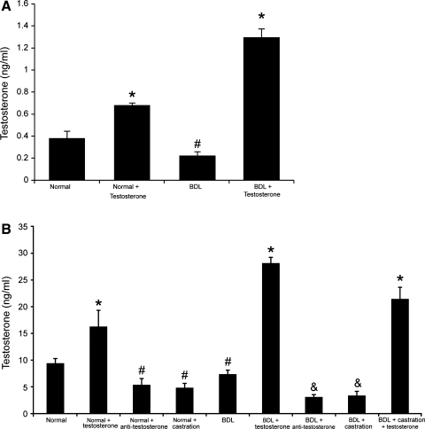
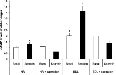
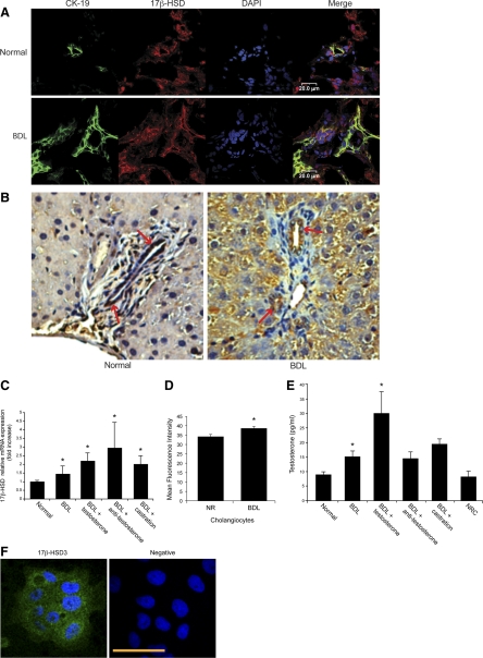
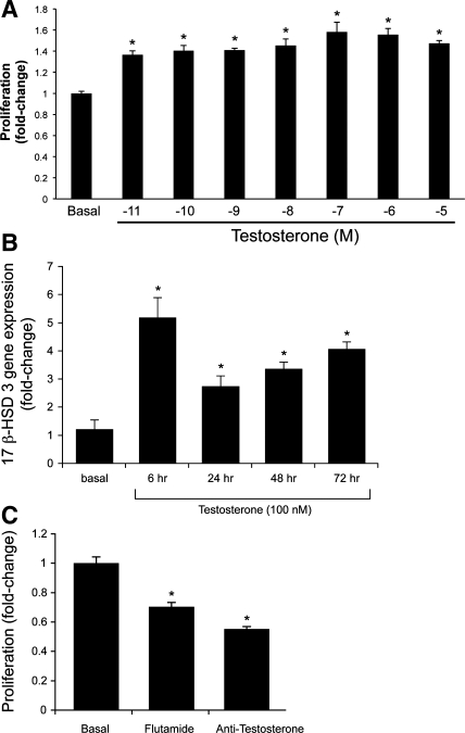
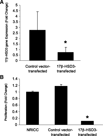
Increased cholangiocyte growth is critical for the maintenance of biliary mass during liver injury by bile duct ligation (BDL). Circulating levels of testosterone decline following castration and during cholestasis. Cholangiocytes secrete sex hormones sustaining cholangiocyte growth by autocrine mechanisms. We tested the hypothesis that testosterone is an autocrine trophic factor stimulating biliary growth. The expression of androgen receptor (AR) was determined in liver sections, male cholangiocytes, and cholangiocyte cultures [normal rat intrahepatic cholangiocyte cultures (NRICC)]. Normal or BDL (immediately after surgery) rats were treated with testosterone or antitestosterone antibody or underwent surgical castration (followed by administration of testosterone) for 1 wk. We evaluated testosterone serum levels; intrahepatic bile duct mass (IBDM) in liver sections of female and male rats following the administration of testosterone; and secretin-stimulated cAMP levels and bile secretion. We evaluated the expression of 17β-hydroxysteroid dehydrogenase 3 (17β-HSD3, the enzyme regulating testosterone synthesis) in cholangiocytes. We evaluated the effect of testosterone on the proliferation of NRICC in the absence/presence of flutamide (AR antagonist) and antitestosterone antibody and the expression of 17β-HSD3. Proliferation of NRICC was evaluated following stable knock down of 17β-HSD3. We found that cholangiocytes and NRICC expressed AR. Testosterone serum levels decreased in castrated rats (prevented by the administration of testosterone) and rats receiving antitestosterone antibody. Castration decreased IBDM and secretin-stimulated cAMP levels and ductal secretion of BDL rats. Testosterone increased 17β-HSD3 expression and proliferation in NRICC that was blocked by flutamide and antitestosterone antibody. Knock down of 17β-HSD3 blocks the proliferation of NRICC. Drug targeting of 17β-HSD3 may be important for managing cholangiopathies.


Figures

Fig. 1.
Fig. 1.
A: image showing that intrahepatic bile ducts from normal and bile duct ligation (BDL) male rats express androgen receptor (AR) (red staining). Colocalization with CK-19 (green staining) of bile ducts expressing the AR (red staining) is also visible. Bar = 50 μm. B: by immunohistochemistry, normal cholangiocytes and hepatocytes express low levels of AR, expression that increased following BDL (A and B; see Table 1). Original magnification, ×40. C: by RT-PCR, the message for AR was expressed by freshly isolated cholangiocytes from normal (NR) and BDL rats and normal rat intrahepatic cholangiocyte cultures (NRICC); the housekeeping gene, glyceraldehyde-3-phosphate dehydrogenase (GAPDH), was expressed similarly by these cells. MW, mol wt. D: by immunofluorescence, NRICC also expressed the protein for AR. Bar = 20 μm. E and F: fluorescence-activated cell sorter (FACS) analysis shows the presence of the protein for AR in freshly isolated cholangiocytes and hepatocytes from normal and BDL male rats; the expression of AR seemed upregulated (*P < 0.05) in BDL cholangiocytes compared with normal cholangiocytes. Data are means ± SE of 3 determinations.
Fig. 2.
Fig. 2.
Measurement of serum testosterone levels in the selected groups of animals. Testosterone serum levels were lower in female and male BDL rats compared with their corresponding normal rats. The administration of testosterone increased testosterone serum levels in normal and BDL female and male rats. Castration significantly decreased testosterone serum levels in normal and BDL rats. Administration of neutralizing antitestosterone antibody decreased testosterone serum levels in both normal and BDL rats compared with rats treated with nonimmune serum. The administration of testosterone to BDL castrated rats partly prevented castration-induced reduction of testosterone serum levels. Data are means ± SE of 12 cumulative determinations. *P < 0.05 compared with the corresponding values of normal rats. #P < 0.05 compared with the corresponding values of normal rats. &P < 0.05 compared with the corresponding values of BDL rats.
Fig. 3.
Fig. 3.
Measurement of basal and secretin-stimulated cAMP levels in purified cholangiocytes. In normal and BDL rats (without castration), secretin increases cAMP levels of purified cholangiocytes. In purified cholangiocytes from normal and BDL castrated rats, there was ablation of the stimulatory effects of secretin on cAMP levels. *P < 0.05 vs. the corresponding basal cAMP levels. #P < 0.05 vs. basal cAMP levels of normal cholangiocytes. Data are means ± SE of 6 experiments.
Fig. 4.
Fig. 4.
A: by immunofluorescence, 17β-hydroxysteroid dehydrogenase 3 (17β-HSD3) was expressed by intrahepatic bile ducts of liver sections from normal and BDL rats with and without castration. Colocalization with CK-19 (green staining) of bile ducts expressing the AR (red staining) is also visible. Bar = 50 μm. B: by immunohistochemistry, cholangiocytes from normal and BDL rats express 17β-HSD3. Original magnification, ×40. The immunoreactivity was higher in bile ducts from male BDL rats (++) compared with their corresponding normal rats (+). C and D: the message and protein for 17β-HSD3 was expressed by normal male cholangiocytes and increased following BDL. Data are means ± SE of 3 real-time PCR and FACS experiments. C: in purified cholangiocytes from BDL rats with castration or receiving antitestosterone antibody, the expression of 17β-HSD3 mRNA was similar to that of BDL cholangiocytes. Data are means ± SE of 3 real-time PCR experiments. *P < 0.05 vs. all of the other groups. D: the protein expression levels of 17β-HSD3 were determined by FACS analysis. *P < 0.05 vs. normal rat. E: normal cholangiocytes and NRICC secrete testosterone in the supernatant. The levels of testosterone increased in the supernatant of BDL cholangiocytes compared with normal cholangiocyte supernatant. In purified cholangiocytes from BDL rats with castration or receiving antitestosterone antibody, the secretion of testosterone was similar to that of BDL cholangiocytes. Data are means ± SE of 8 evaluations. *P < 0.05 vs. all of the other groups. F: by immunofluorescence, NRICC express the protein for 17β-HSD3. Bar = 50 μm.
Fig. 5.
Fig. 5.
A: measurement of NRICC proliferation following treatment with 0.2% BSA or testosterone. Testosterone (10−11 to 10−5 M) in vitro increased the proliferation (by MTS) of NRICC compared with the corresponding basal value. Data are means ± SE of 6 experiments. *P < 0.05 vs. the corresponding basal value. B: testosterone in vitro increased the mRNA expression of 17β-HSD3 (the key enzyme regulating testosterone synthesis) in NRICC compared with the corresponding basal value. Data are means ± SE of 6 experiments. *P < 0.05 vs. the corresponding basal value. C: measurement of cell growth (by MTS assays) in NRICC stimulated with BSA (basal), flutamide (a specific antagonist of AR), or antitestosterone antibody for 48 h. When NRICC were incubated with flutamide or antitestosterone antibody, there was a decrease in cell growth compared with NRICC treated with BSA. Data are means ± SE of 6 experiments. *P < 0.05 vs. the corresponding basal value.
Fig. 6.
Fig. 6.
A and B: effect of knock down of 17β-HSD3 on the basal proliferative activity of NRICC by MTS assays. Knock down of 17β-HSD3 (∼80%) in NRICC (A) decreased the proliferative activity of NRICC (B), supporting the concept that testosterone is an autocrine factor sustaining biliary growth. Data are means ± SE of 6 experiments. *P < 0.05 vs. the corresponding basal value.

Similar articles

Cited by

References

    1. Alpini G, Glaser S, Robertson W, Phinizy JL, Rodgers RE, Caligiuri A, LeSage G. Bile acids stimulate proliferative and secretory events in large but not small cholangiocytes. Am J Physiol Gastrointest Liver Physiol 273: G518– G529, 1997 - PubMed
    1. Alpini G, Glaser S, Ueno Y, Pham L, Podila PV, Caligiuri A, LeSage G, LaRusso NF. Heterogeneity of the proliferative capacity of rat cholangiocytes after bile duct ligation. Am J Physiol Gastrointest Liver Physiol 274: G767– G775, 1998 - PubMed
    1. Alpini G, Lenzi R, Sarkozi L, Tavoloni N. Biliary physiology in rats with bile ductular cell hyperplasia. Evidence for a secretory function of proliferated bile ductules. J Clin Invest 81: 569– 578, 1988 - PMC - PubMed
    1. Alpini G, Phinizy JL, Glaser S, Francis H, Benedetti A, Marucci L, LeSage G. Development and characterization of secretin-stimulated secretion of cultured rat cholangiocytes. Am J Physiol Gastrointest Liver Physiol 284: G1066– G1073, 2003 - PubMed
    1. Alpini G, Prall RT, LaRusso NF. The pathobiology of biliary epithelia. The Liver; Biology & Pathobiology (4th ed.), edited by Arias IM, Boyer JL, Chisari FV, Fausto N, Jakoby W, Schachter D, Shafritz DA. Philadelphia, PA: Williams & Wilkins, 2001, p. 421–435

Publication types

MeSH terms