

Image quality and radiation dose of dual-energy CT of the head and neck compared with a standard 120-kVp acquisition
- PMID: 21903909
- PMCID: PMC7964383
- DOI: 10.3174/ajnr.A2654
Image quality and radiation dose of dual-energy CT of the head and neck compared with a standard 120-kVp acquisition
Abstract
Background and purpose: DECT offers additional image datasets with potential benefits, but its use for H&N imaging is not justified unless image quality is preserved without increased radiation dose. The aim of this work was to compare image quality and radiation dose between a DE-derived WA image dataset and a standard SECT acquisition of the H&N.
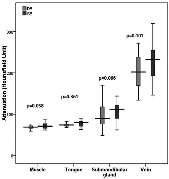
Materials and methods: Thirty-two patients underwent DECT of the H&N (tube voltages 80 and Sn140 kVp) and were compared with the last 32 patients who underwent standard SECT (120 kVp) on the same dual-source scanner. WA images from the 2 DE tubes were compared with images obtained with an SE mode. Radiation doses and attenuation measurements of the internal jugular vein, submandibular gland, and sternomastoid and tongue muscles were compared. Objective image noise was compared at 5 anatomic levels. Two blinded readers compared subjective image quality by using 5-point grading scales.
Results: CTDI(vol) was 12% lower with DE than with SECT, a difference of 1.5 mGy, (P < .0001). Objective noise was not significantly different between DE and SECT at any of the anatomic levels (P > .05). No significant differences in attenuation measurements were observed between DE and SECT (P > .05). No significant differences in subjective image quality scores were observed between DE and SECT at any of the 5 anatomic levels (P > .05).
Conclusions: DE-derived WA images of the H&N are equivalent to standard SE acquisitions and thus can be used for routine diagnostic purposes. Multiple additional image datasets can be obtained with no radiation dose penalty.
Figures


References
-
- Kang MJ, Park CM, Lee CH, et al. Dual-energy CT: clinical applications in various pulmonary diseases. Radiographics 2010;30:685–98 - PubMed
-
- Johnson TR, Krauss B, Sedlmair M, et al. Material differentiation by dual energy CT: initial experience. Eur Radiol 2007;17:1510–17 - PubMed
-
- Stolzmann P, Leschka S, Scheffel H, et al. Characterization of urinary stones with dual-energy CT: improved differentiation using a tin filter. Invest Radiol 2010;45:1–6 - PubMed
-
- Thieme SF, Johnson TR, Lee C, et al. Dual-energy CT for the assessment of contrast material distribution in the pulmonary parenchyma. AJR Am J Roentgenol 2009;193:144–49 - PubMed
-
- Morhard D, Fink C, Graser A, et al. Cervical and cranial computed tomographic angiography with automated bone removal: dual energy computed tomography versus standard computed tomography. Invest Radiol 2009;44:293–97 - PubMed
Publication types
MeSH terms
LinkOut - more resources
Full Text Sources
Medical