

Therapeutic modulation of cerebral microhemorrhage in a mouse model of cerebral amyloid angiopathy
- PMID: 21903962
- PMCID: PMC3221609
- DOI: 10.1161/STROKEAHA.111.626655
Therapeutic modulation of cerebral microhemorrhage in a mouse model of cerebral amyloid angiopathy
Abstract
Background and purpose: The aging brain demonstrates frequent MRI and pathological evidence of cerebral microbleeds, which are often associated with cerebral amyloid angiopathy. To develop new therapeutic strategies for this disorder, we studied cerebral microhemorrhage in a well-characterized mouse model of cerebral amyloid angiopathy.
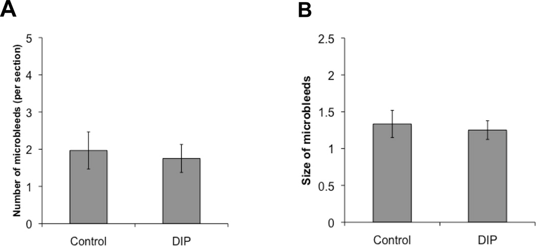
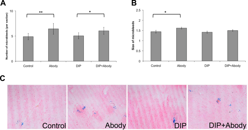
Methods: Tg2576 mice were studied at ages ranging from 2 to 21 months. Spontaneous and induced microscopic bleeding was analyzed with and without a passive anti-amyloid immunization regimen and dietary supplementation of ischemic stroke prevention medication dipyridamole.
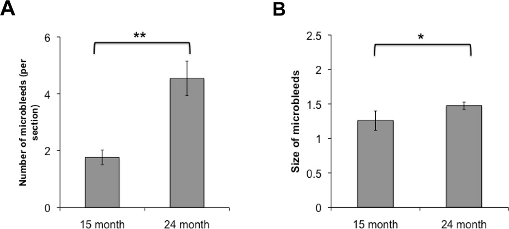
Results: Areas of microhemorrhage were easily demonstrated and were significantly more prominent in the oldest mice and in animals treated with anti-amyloid immunotherapy. Dipyridamole supplementation in the diet generated plasma levels >790 ng/mL within the range seen clinically. Dipyridamole treatment did not worsen frequency and size of cerebral microscopic bleeding.
Conclusions: The Tg2576 mouse is a useful model to study progression and modification of spontaneous and immunotherapy-induced cerebral microhemorrhage. Absence of microhemorrhage worsening with dipyridamole treatment suggests a potential therapeutic role of this agent when ischemic and microhemorrhagic lesions coexist.
Figures
References
-
- Vernooij MW, van der Lugt A, Ikram MA, Wielopolski PA, Niessen WJ, Hofman A, et al. Prevalence and risk factors of cerebral microbleeds: the Rotterdam Scan Study. Neurology. 2008;70:1208–1214. - PubMed
-
- Cullen KM, Kocsi Z, Stone J. Pericapillary haem-rich deposits: evidence for microhaemorrhages in aging human cerebral cortex. J Cereb Blood Flow Metab. 2005;25:1656–1667. - PubMed
-
- Young VG, Halliday GM, Kril JJ. Neuropathologic correlates of white matter hyperintensities. Neurology. 2008;71:804–811. - PubMed
Publication types
MeSH terms
Substances
Grants and funding
- T34 GM069337/GM/NIGMS NIH HHS/United States
- P01 AG000538/AG/NIA NIH HHS/United States
- NS050895/NS/NINDS NIH HHS/United States
- P50 AG016573/AG/NIA NIH HHS/United States
- R01 NS050895/NS/NINDS NIH HHS/United States
- AG00538/AG/NIA NIH HHS/United States
- P50AG16573/AG/NIA NIH HHS/United States
- R01 AG020241/AG/NIA NIH HHS/United States
- R01 NS020989/NS/NINDS NIH HHS/United States
- AG020241/AG/NIA NIH HHS/United States
- GM-69337/GM/NIGMS NIH HHS/United States
- RF1 AG020241/AG/NIA NIH HHS/United States
- NS20989/NS/NINDS NIH HHS/United States
LinkOut - more resources
Full Text Sources
Other Literature Sources
Medical
