. 2011 Sep 1;67(Pt 9):1123-8.
doi: 10.1107/S174430911102690X. Epub 2011 Aug 16.

Structure of fumarate hydratase from Rickettsia prowazekii, the agent of typhus and suspected relative of the mitochondria

Affiliations

Structure of fumarate hydratase from Rickettsia prowazekii, the agent of typhus and suspected relative of the mitochondria

Isabelle Phan et al. Acta Crystallogr Sect F Struct Biol Cryst Commun. .

Abstract

Rickettsiae are obligate intracellular parasites of eukaryotic cells that are the causative agents responsible for spotted fever and typhus. Their small genome (about 800 protein-coding genes) is highly conserved across species and has been postulated as the ancestor of the mitochondria. No genes that are required for glycolysis are found in the Rickettsia prowazekii or mitochondrial genomes, but a complete set of genes encoding components of the tricarboxylic acid cycle and the respiratory-chain complex is found in both. A 2.4 Å resolution crystal structure of R. prowazekii fumarate hydratase, an enzyme catalyzing the third step of the tricarboxylic acid cycle pathway that ultimately converts phosphoenolpyruvate into succinyl-CoA, has been solved. A structure alignment with human mitochondrial fumarate hydratase highlights the close similarity between R. prowazekii and mitochondrial enzymes.


Figures

Figure 1
Figure 1
Chemical reaction pathway of the tricarboxylic acid cycle (TCA; also known as the Krebs cycle and the citric acid cycle); catalytic enzymes are indicated in pink boxes, with fumarase, the subject of this study, highlighted in red. This figure was prepared with CellDesigner (Funahashi et al., 2003 ▶).
Figure 2
Figure 2
Ribbon diagram of the homodimeric unit structure of R. prowazekii FumC showing (a) the overall fold gradient-coloured from red (N-terminus) to blue (C-­terminus) and (b) the backbone trace of chain A (blue) and chain B (brown) of the dimeric unit. This figure and all other structure figures in this paper (except for Fig. 5 ▶) were prepared using the POV-Ray renderer (http://povray.org) and DeepView (Guex & Peitsch, 1997 ▶).
Figure 3
Figure 3
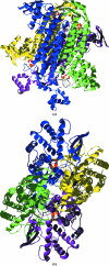
Two views of the tetrameric assembly predicted by PISA from the 3gtd coordinates (http://www.ebi.ac.uk/msd-srv/prot_int/pistart.html) showing the schematic backbone trace of the four subunits modelled for dimer 1 chain A (blue) and B (yellow) and dimer 2 chain A (magenta) and B (green). (a) The side view of each chain bound to the ligand malonate shown in CPK. (b) The two sodium ions at the interface of each dimer can be seen near the central axis of symmetry.
Figure 4
Figure 4
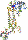
Superposition of the backbone Cα traces of the FumC monomers from Rickettsia (PDB entry 3gtd; red), human (PDB entry 3e04; green; Structural Genomics Consortium, unpublished work), E. coli (PDB entry 1kq7; blue; Estévez et al., 2002 ▶) and S. cerevisiae (PDB entry 1yfm; yellow; Weaver et al., 1998 ▶) showing the conserved overall fold and the deviations at the C-terminus at the bottom left region of the structure. The ligands for the Rickettsia and E. coli structures, malonate (red) and citric acid (blue), respectively, are represented in CPK colors.
Figure 5
Figure 5
Schematic representation of the ligand environment in the Rickettsia FumC monomer complexed with malonate superimposed on the corresponding residues in the human structure (PDB entry 3e04). The backbone (gray for Rickettsia, magenta for human) and side chains of residues located within 6 Å of the ligand are shown. Hydrogen bonds of less than 3 Å are shown as dashed lines. Residues are numbered according to the Rickettsia FumC numbering system and residues from the second protomer that comprise the active site are identified with the ′ notation (Thr187′ and His188′). The 2|F o| − |F c| electron-density map is shown in light blue mesh contoured at 1.0σ. Note that the human structure is apo and thus Lys371 (equivalent to Lys324 in Rickettsia) appears disordered. This figure was generated using PyMOL (DeLano, 2002 ▶).

Similar articles

Cited by

References

    1. Andersson, S. G., Zomorodipour, A., Andersson, J. O., Sicheritz-Pontén, T., Alsmark, U. C., Podowski, R. M., Näslund, A. K., Eriksson, A. S., Winkler, H. H. & Kurland, C. G. (1998). Nature (London), 396, 133–140. - PubMed
    1. Aslanidis, C. & de Jong, P. J. (1990). Nucleic Acids Res. 18, 6069–6074. - PMC - PubMed
    1. Azad, A. F. (2007). Clin. Infect. Dis. 45, S52–S55. - PubMed
    1. Bayley, J. P., Launonen, V. & Tomlinson, I. P. (2008). BMC Med. Genet. 9, 20. - PMC - PubMed
    1. Chen, V. B., Arendall, W. B., Headd, J. J., Keedy, D. A., Immormino, R. M., Kapral, G. J., Murray, L. W., Richardson, J. S. & Richardson, D. C. (2010). Acta Cryst. D66, 12–21. - PMC - PubMed

Publication types

MeSH terms

Substances

Associated data