. 2011 Sep 1;67(Pt 9):1129-36.
doi: 10.1107/S1744309111006336. Epub 2011 Aug 16.

Solution structure of an arsenate reductase-related protein, YffB, from Brucella melitensis, the etiological agent responsible for brucellosis

Affiliations

Solution structure of an arsenate reductase-related protein, YffB, from Brucella melitensis, the etiological agent responsible for brucellosis

Garry W Buchko et al. Acta Crystallogr Sect F Struct Biol Cryst Commun. .

Abstract

Brucella melitensis is the etiological agent responsible for brucellosis. Present in the B. melitensis genome is a 116-residue protein related to arsenate reductases (Bm-YffB; BR0369). Arsenate reductases (ArsC) convert arsenate ion (H(2)AsO(4)(-)), a compound that is toxic to bacteria, to arsenite ion (AsO(2)(-)), a product that may be efficiently exported out of the cell. Consequently, Bm-YffB is a potential drug target because if arsenate reduction is the protein's major biological function then disabling the cell's ability to reduce arsenate would make these cells more sensitive to the deleterious effects of arsenate. Size-exclusion chromatography and NMR spectroscopy indicate that Bm-YffB is a monomer in solution. The solution structure of Bm-YffB (PDB entry 2kok) shows that the protein consists of two domains: a four-stranded mixed β-sheet flanked by two α-helices on one side and an α-helical bundle. The α/β domain is characteristic of the fold of thioredoxin-like proteins and the overall structure is generally similar to those of known arsenate reductases despite the marginal sequence similarity. Chemical shift perturbation studies with (15)N-labeled Bm-YffB show that the protein binds reduced glutathione at a site adjacent to a region similar to the HX(3)CX(3)R catalytic sequence motif that is important for arsenic detoxification activity in the classical arsenate-reductase family of proteins. The latter observation supports the hypothesis that the ArsC-YffB family of proteins may function as glutathione-dependent thiol reductases. However, comparison of the structure of Bm-YffB with the structures of proteins from the classical ArsC family suggest that the mechanism and possibly the function of Bm-YffB and other related proteins (ArsC-YffB) may differ from those of the ArsC family of proteins.


Figures

Figure 1
Figure 1
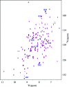
Assigned 1H–15N HSQC spectrum of double-labeled Bm-YffB collected at 293 K in NMR buffer (100 mM NaCl, 20 mM Tris–HCl, 1.0 mM DTT, pH 7.1) at a 1H resonance frequency of 750 MHz. Side-chain –NH2 resonances are indicated by dashed horizontal lines (red) and the exchangeable ring resonances for Trp20 and Trp53 are identified with an ‘r’.
Figure 2
Figure 2
(a) Superposition of the cartoon representations of the ensemble of structures calculated for Bm-YffB (PDB entry 2kok), with α-helices colored blue and β-strands colored gold. (b) Cartoon representation of the structure most similar to the average structure of the ensemble, with the four β-strands and seven α-helices labeled.
Figure 3
Figure 3
Stereoview showing a cartoon representation of the structure most similar to the average structure in the ensemble, with the protein rainbow-colored (ROYGBIV) from the N-terminus to the C-terminus.
Figure 4
Figure 4
Maps generated using PyMOL (DeLano, 2002 ▶) of the electrostatic potential at the solvent-accessible surface of Bm-YffB. The long axis of the protein is illustrated and is sequentially rotated 90° about the horizontal axis four times.
Figure 5
Figure 5
(a) Circular dichroism steady-state wavelength spectrum for Bm-YffB (0.05 mM) in NMR buffer collected at 298 K. (b) The CD thermal melt for Bm-YffB obtained by measuring the ellipticity at 216 nm in 2.0 K intervals between 283 and 353 K. (c) The first derivative of the thermal melt curve shows that the protein has a melting temperature of 326.6 K.
Figure 6
Figure 6
Superposition of the structure closest to the mean for Bm-YffB (PDB entry 2kok, blue) on the crystal structure determined for Pa-YffB (PDB entry 1rw1, magenta; Teplyakov et al., 2004 ▶) using the program SuperPose (Maiti et al., 2004 ▶).
Figure 7
Figure 7
Superposition of the structure closest to the mean for Bm-YffB (PDB entry 2kok, blue) on the crystal structure determined for E. coli ArsC (PDB entry 1id9, magenta; Martin et al., 2001 ▶) using the program SuperPose (Maiti et al., 2004 ▶). The side chains of the three arginine residues in Ec-ArsC (Arg60, Arg94 and Arg107) that form a thiasahydroxyl adduct that is essential for enzymatic function are labeled and colored red. The only residue in Bm-YffB that is equivalent to those in the arginine triad of Ec-ArsC is Arg96; this side chain is colored black.
Figure 8
Figure 8
Overlay of the 1H–15N HSQC spectrum of 15N-labeled Bm-YffB (black) on the spectrum collected in the presence of a 2:1 molar ratio of glutathione:Bm-YffB (magenta). Residues that were significantly perturbed are labeled. The spectrum at an ∼1:1 molar ratio of glutathione:Bm-YffB was similar to that shown here at a 2:1 molar ratio. Data were collected at a proton resonance frequency of 600 MHz at 293 K.
Figure 9
Figure 9
Surface representation of the structure of Bm-YffB shown with a labeled transparent cartoon representation. Regions that may potentially be important for the function of the protein are labeled. The lone equivalent residue to those present in the arginine triad of Ec-ArsC, Arg96, is colored black, residues in the potential GX 3CX 3K catalytic sequence motif are colored yellow and the residues perturbed by the addition of reduced glutathione (labeled in Fig. 8 ▶) are colored red.

Similar articles

Cited by

References

    1. Bhattacharjya, S., Xu, P., Gingras, R., Shaykhutdinov, R., Wu, C., Whiteway, M. & Ni, F. (2004). J. Mol. Biol. 344, 1071–1087. - PubMed
    1. Bhattacharya, A., Tejero, R. & Montelione, G. T. (2007). Proteins, 66, 778–795. - PubMed
    1. Bryan, C. M., Bhandari, J., Napuli, A. J., Leibly, D. J., Choi, R., Kelley, A., Van Voorhis, W. C., Edwards, T. E. & Stewart, L. J. (2011). Acta Cryst. F67, 1010–1014. - PMC - PubMed
    1. Buchko, G. W., Daughdrill, G. W., de Lorimier, R., Rao, B. K., Isern, N. G., Lingbeck, J. M., Taylor, J. S., Wold, M. S., Gochin, M., Spicer, L. D., Lowry, D. F. & Kennedy, M. A. (1999). Biochemistry, 38, 15116–15128. - PMC - PubMed
    1. Buchko, G. W., Tarasevich, B. J., Bekhazi, J., Snead, M. L. & Shaw, W. J. (2008). Biochemistry, 47, 13215–13222. - PMC - PubMed

Publication types

MeSH terms

Substances

Associated data