. 2011 Sep 1;67(Pt 9):1154-8.
doi: 10.1107/S1744309111029575. Epub 2011 Aug 16.

Structure of the cystathionine γ-synthase MetB from Mycobacterium ulcerans

Affiliations

Structure of the cystathionine γ-synthase MetB from Mycobacterium ulcerans

Matthew C Clifton et al. Acta Crystallogr Sect F Struct Biol Cryst Commun. .

Abstract

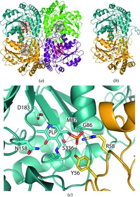
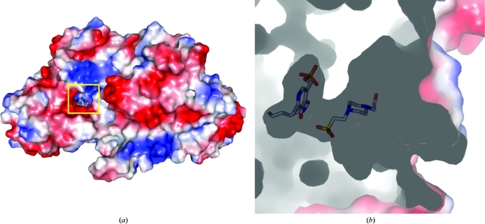
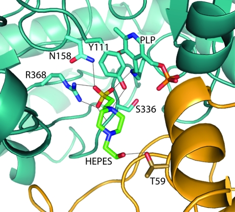
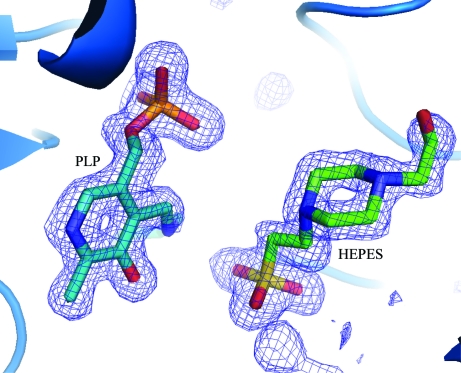
Cystathionine γ-synthase (CGS) is a transulfurication enzyme that catalyzes the first specific step in L-methionine biosynthesis by the reaction of O(4)-succinyl-L-homoserine and L-cysteine to produce L-cystathionine and succinate. Controlling the first step in L-methionine biosythesis, CGS is an excellent potential drug target. Mycobacterium ulcerans is a slow-growing mycobacterium that is the third most common form of mycobacterial infection, mainly infecting people in Africa, Australia and Southeast Asia. Infected patients display a variety of skin ailments ranging from indolent non-ulcerated lesions as well as ulcerated lesions. Here, the crystal structure of CGS from M. ulcerans covalently linked to the cofactor pyridoxal phosphate (PLP) is reported at 1.9 Å resolution. A second structure contains PLP as well as a highly ordered HEPES molecule in the active site acting as a pseudo-ligand. These results present the first structure of a CGS from a mycobacterium and allow comparison with other CGS enzymes. This is also the first structure reported from the pathogen M. ulcerans.


Figures

Figure 1
Figure 1
The overall structure of MetB from M. ulcerans. (a) In both the PLP and the PLP and HEPES complex structures a tetramer was found similar to previous MetB structures. Each tetramer contains two functional dimers (yellow/blue, purple/green). Active sites facing out of the page are indicated in black, while active sites facing into the page are indicated in red. (b) An individual dimer of MetB contains two active sites. The main portion of the active site is made up by a pocket within the individual monomer, with a flexible loop from the second monomer closing the pocket. The PLP moiety is shown in gray. (c) The active site of MetB covalently linked to PLP. A tight hydrogen-bond network is created around the PLP moiety by residues from both of the individual monomers in the homodimer. Monomer A is indicated in blue and monomer B in yellow; hydrogen bonds are represented by black dashed lines and range from 2.4 to 3.2 Å in length. The average B factor for PLP for the MetB–PLP structure is 14.2 Å2.
Figure 2
Figure 2
A surface electrostatics representation of MetB–PLP–HEPES. (a) The surface shows a small pocket that forms the active site between the individual monomers. (b) A side view of the surface representation shows PLP buried deep in the active site and HEPES bound tightly in the position expected for the starting reactant O 4-succinyl-l-­homoserine.
Figure 3
Figure 3
The active site of MetB covalently linked to PLP and bound to HEPES. The tight binding environment for the HEPES molecule is formed by a hydrogen-bonding network created by molecules of both monomers in the homodimer. Monomer A is indicated in blue, monomer B in yellow and HEPES in green; hydrogen bonds are represented by black dashed lines and range from 2.4 to 3.2 Å in length. The average B factors for the PLP and HEPES molecules in the MetB–PLP–HEPES structure are 9.1 and 15.9 Å2, respectively.
Figure 4
Figure 4
Electron density in the active site of MetB–PLP–HEPES. A 2F oF c electron-density map contoured at 3σ (blue) clearly shows the positions of both PLP and HEPES in the binding site of MetB.

Similar articles

Cited by

References

    1. Alexandrov, A., Dutta, K. & Pascal, S. M. (2001). Biotechniques, 30, 1194–1198. - PubMed
    1. Alexandrov, A., Vignali, M., LaCount, D. J., Quartley, E., de Vries, C., De Rosa, D., Babulski, J., Mitchell, S. F., Schoenfeld, L. W., Fields, S., Hol, W. G., Dumont, M. E., Phizicky, E. M. & Grayhack, E. J. (2004). Mol. Cell. Proteomics, 3, 934–938. - PubMed
    1. Amadasi, A., Bertoldi, M., Contestabile, R., Bettati, S., Cellini, B., di Salvo, M. L., Borri-Voltattorni, C., Bossa, F. & Mozzarelli, A. (2007). Curr. Med. Chem. 14, 1291–1324. - PubMed
    1. Aslanidis, C. & de Jong, P. J. (1990). Nucleic Acids Res. 18, 6069–6074. - PMC - PubMed
    1. Chen, V. B., Arendall, W. B., Headd, J. J., Keedy, D. A., Immormino, R. M., Kapral, G. J., Murray, L. W., Richardson, J. S. & Richardson, D. C. (2010). Acta Cryst. D66, 12–21. - PMC - PubMed

Publication types

MeSH terms

Substances