. 2011 Oct;34(8):597-605.
doi: 10.1097/CJI.0b013e3182307fd8.

Redirected lysis of human melanoma cells by a MCSP/CD3-bispecific BiTE antibody that engages patient-derived T cells

Affiliations

Redirected lysis of human melanoma cells by a MCSP/CD3-bispecific BiTE antibody that engages patient-derived T cells

Hitoe Torisu-Itakura et al. J Immunother. 2011 Oct.

Abstract

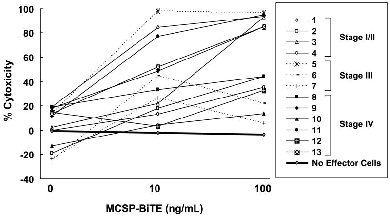
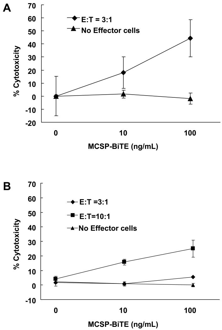
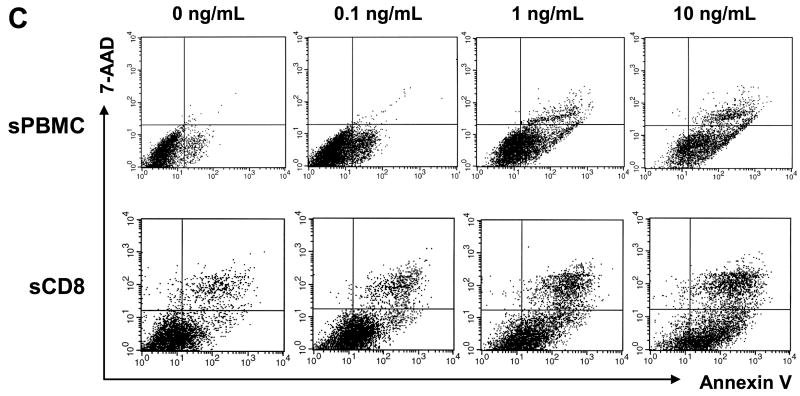
Melanoma-associated chondroitin sulfate proteoglycan (MCSP; also called HMW-MAA, CSPG4, NG2, MSK16, MCSPG, MEL-CSPG, or gp240) is a well characterized melanoma cell-surface antigen. In this study, a new bispecific T-cell engaging (BiTE) antibody that binds to MCSP and human CD3 (MCSP-BiTE) was tested for its cytotoxic activity against human melanoma cell lines. When unstimulated peripheral mononuclear blood cells (PBMCs) derived from healthy donors were cocultured with melanoma cells at effector:target ratios of 1:1, 1:5, or 1:10, and treated with MCSP-BiTE antibody at doses of 10, 100, or 1000 ng/mL, all MCSP-expressing melanoma cell lines (n=23) were lysed in a dose-dependent and effector:target ratio-dependent manner, whereas there was no cytotoxic activity against MCSP-negative melanoma cell lines (n=2). To investigate whether T cells from melanoma patients could act as effector cells, we cocultured unstimulated PBMCs with allogeneic melanoma cells from 13 patients (4 stage I/II, 3 stage III, and 6 stage IV) or with autologous melanoma cells from 2 patients (stage IV). Although cytotoxic activity varied, all 15 PBMC samples mediated significant redirected lysis by the BiTE antibody. When PBMC or CD8 T cells were prestimulated by anti-CD3 antibody OKT-3 and interleukin-2, the MCSP-BiTE concentrations needed for melanoma cell lysis decreased up to 1000-fold. As MCSP is expressed on most human melanomas, immunotherapy with MCSP/CD3-bispecific antibodies merits clinical investigation.


Figures

Figure 1
Figure 1
Cytotoxicity of MCSP-BiTE antibody treatment in human melanoma cell lines. Twenty-three MCSP-positive (1A) or 2 MCSP-negative melanoma cell lines (1B) were co-cultured with effector cells (PBMC obtained from healthy donors) at E:T ratios of 1:10, 1:5, or 1:1. Cells were then treated with MCSP-BiTE antibody in concentrations of 0, 10, 100 or 1,000 ng/mL. Percent change in cytotoxicity from baseline (MCSP-BiTE 0 ng/mL) is shown as the mean and standard error (SE) of triplicate determinations. C: Means and SE of 23 MCSP-positive melanoma cell lines. Number of live melanoma cells after co-culture with or without effector cells at E:T ratios of 1:10, 1:5 and 1:1, and treatment with 0 or 100 ng/mL of MCSP-BiTE. The anti-tumor effect of MCSP-BiTE was calculated according to the following formula: live melanoma cell number = total melanoma cell counts (PKH67 positive cells) × percentage of live melanoma cell (PI negative cells).
Figure 1
Figure 1
Cytotoxicity of MCSP-BiTE antibody treatment in human melanoma cell lines. Twenty-three MCSP-positive (1A) or 2 MCSP-negative melanoma cell lines (1B) were co-cultured with effector cells (PBMC obtained from healthy donors) at E:T ratios of 1:10, 1:5, or 1:1. Cells were then treated with MCSP-BiTE antibody in concentrations of 0, 10, 100 or 1,000 ng/mL. Percent change in cytotoxicity from baseline (MCSP-BiTE 0 ng/mL) is shown as the mean and standard error (SE) of triplicate determinations. C: Means and SE of 23 MCSP-positive melanoma cell lines. Number of live melanoma cells after co-culture with or without effector cells at E:T ratios of 1:10, 1:5 and 1:1, and treatment with 0 or 100 ng/mL of MCSP-BiTE. The anti-tumor effect of MCSP-BiTE was calculated according to the following formula: live melanoma cell number = total melanoma cell counts (PKH67 positive cells) × percentage of live melanoma cell (PI negative cells).
Figure 2
Figure 2
Anti-tumor effects of MCSP-BiTE antibody with melanoma patients' PBMC against the MCSP-positive melanoma cell line (M27-HI). PBMC from 13 melanoma patients were cocultured with M27-HI at an E:T ratio 3:1; cells were treated with MCSP-BiTE antibody (0, 10 or 100 ng/mL) or control BiTE (100 ng/mL). The results of control BiTE that binds CD3 but not MCSP were used as baseline data. The numbers 1-13 correspond to the patient numbers in column 1 of Table 1.
Figure 3
Figure 3
Anti-tumor effects of MCSP-BiTE antibody using MCSP-positive autologous melanoma cells and PBMC. Cells were treated with MCSP-BiTE antibody (0, 10 or 100 ng/mL) or control BiTE (100 ng/mL). Means from triplicate determination and SE are shown. A: Autologous PBMC for M34-HI melanoma cell line was cocultured with M34-HI melanoma cell line at an E:T ratio 3:1. B: Autologous PBMC for M35-HI melanoma cell line was cocultured with M35-HI cell line at an E:T ratio 3:1 and 10:1.
Figure 4
Figure 4
Use of OKT-3 and IL-2 to stimulate PBMC or CD8+ T cells from healthy donors increased the efficacy of MCSP-BiTE redirected lysis in MCSP-positive melanoma cell (M11-HI). A: Percent change of cytotoxicity in M11-HI cell line co-cultured with stimulated PBMC (sPBMC) or CD8+ T cells (sCD8) from healthy donors at E:T ratio 1:1. Value of MCSP-BiTE 0 ng/mL was used as a baseline in stimulated PBMC and CD8+ T cells, respectively. B: Number of live melanoma cells after FACS-based cytotoxicity assay. The anti-tumor effect of MCSP-BiTE was calculated according to the following formula: live melanoma cell number = total melanoma cell number (PKH67 positive) × percentage of live melanoma cell (7-AAD negative and annexin V negative). All data from 4 sets of PBMC or CD8+ T cells co-cultured with M11-HI was averaged. Each bar shows the mean number of live melanoma cells and SE. C: Melanoma cells were pre-stained with PKH-67 and co-cultured with stimulated PBMC or CD8+ T cells for 18 hours in presence of MCSP-BiTE 0, 0.1, 1 and 10 ng/mL. 7-AAD and annexin V were used to detect dead melanoma cells and early apoptotic cells, respectively. sPBMC: stimulated PBMC, sCD8: stimulated CD8+ T cell.
Figure 4
Figure 4
Use of OKT-3 and IL-2 to stimulate PBMC or CD8+ T cells from healthy donors increased the efficacy of MCSP-BiTE redirected lysis in MCSP-positive melanoma cell (M11-HI). A: Percent change of cytotoxicity in M11-HI cell line co-cultured with stimulated PBMC (sPBMC) or CD8+ T cells (sCD8) from healthy donors at E:T ratio 1:1. Value of MCSP-BiTE 0 ng/mL was used as a baseline in stimulated PBMC and CD8+ T cells, respectively. B: Number of live melanoma cells after FACS-based cytotoxicity assay. The anti-tumor effect of MCSP-BiTE was calculated according to the following formula: live melanoma cell number = total melanoma cell number (PKH67 positive) × percentage of live melanoma cell (7-AAD negative and annexin V negative). All data from 4 sets of PBMC or CD8+ T cells co-cultured with M11-HI was averaged. Each bar shows the mean number of live melanoma cells and SE. C: Melanoma cells were pre-stained with PKH-67 and co-cultured with stimulated PBMC or CD8+ T cells for 18 hours in presence of MCSP-BiTE 0, 0.1, 1 and 10 ng/mL. 7-AAD and annexin V were used to detect dead melanoma cells and early apoptotic cells, respectively. sPBMC: stimulated PBMC, sCD8: stimulated CD8+ T cell.

References

    1. Baeuerle PA, Kufer P, Bargou R. BiTE: Teaching antibodies to engage T-cells for cancer therapy. Curr Opin Mol Ther. 2009;11:22–30. - PubMed
    1. Baeuerle PA, Reinhardt C. Bispecific T-cell engaging antibodies for cancer therapy. Cancer Res. 2009;69:4941–4944. - PubMed
    1. Bargou R, Leo E, Zugmaier G, et al. Tumor regression in cancer patients by very low doses of a T cell-engaging antibody. Science. 2008;321:974–977. - PubMed
    1. Brischwein K, Parr L, Pflanz S, et al. Strictly target cell-dependent activation of T cells by bispecific single-chain antibody constructs of the BiTE class. J Immunother. 2007;30:798–807. - PubMed
    1. Haas C, Krinner E, Brischwein K, et al. Mode of cytotoxic action of T cell-engaging BiTE antibody MT110. Immunobiology. 2009;214:441–453. - PubMed

Publication types