Review
. 2011 Sep 9;11(10):672-84.
doi: 10.1038/nri3066.

New insights into the T cell synapse from single molecule techniques

Affiliations
Review

New insights into the T cell synapse from single molecule techniques

Michael L Dustin et al. Nat Rev Immunol. .

Abstract

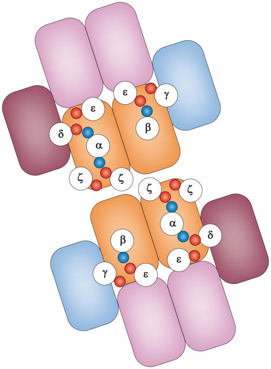
T cell activation depends on extracellular ligation of the T cell receptor (TCR) by peptide-MHC complexes in a synapse between the T cell and an antigen-presenting cell. The process then requires the assembly of signalling complexes between the TCR and the adaptor protein linker for activation of T cells (LAT), and subsequent filamentous actin (F-actin)-dependent TCR cluster formation. Recent progress in each of these areas, made possible by the emergence of new techniques, has forced us to rethink our assumptions and consider some radical new models. These describe the receptor interaction parameters that control T cell responses and the mechanism by which LAT is recruited to the TCR signalling machinery. This is an exciting time in T cell biology, and further innovation in imaging and genomics is likely to lead to a greater understanding of how T cells are activated.


Figures

Figure 1
Figure 1
A The three layers on the T cell side of the immunological synapse are pictured here : receptor layer (TCR complex, CD4/CD8, CD28, LFA1), signalling layer (LCK, ZAP70, ITK, PLCγ) and the cytoskeleton layer (talin, vinculin, FAK/PYK2, paxillin and actin). The adaptor protein LAT is shown attached to the plasma membrane and anchored to vesicles as discussed in the text. B. The supramolecular clusters of an immunological synapse. TCR/cSMAC core (red), CD28/cSMAC periphery (green) and LFA-1/pSMAC (purple). The entire contact area is outlined, and the yellow color represents f-actin, which forms dynamic protrusions that move around the cell periphery (dSMAC) in a radial wave. Each SMAC contains hundreds to thousands of receptors and the dSMAC is the site in which TCR microclusters are first detected.
Figure 2
Figure 2
Asymmetry of CD3 complexes exposes a dimerisation interface. Schematic top-down view of αβ TCR dimer proposed by Kuhns et al. Outlined circles represent the indicated transmembrane helices based on distances taken from the ζζ dimer NMR structure. The small blue (positive) and red (negative) circles represent the intermembrane charged residues. The boxes schematically represent the Ig-like domains of α (light blue), β (medium blue), γ (blue-green), δ (olive}. and ε (purple).
Figure 3
Figure 3
Two views of TCR and LAT clusters. A and C. Dynamic protein ‘islands‘. TCR complexes in red and LAT molecules in green are organized in concatemers within protein islands in the steady state. After peptide–MHC complex recognition (not represented) the concatemers fuse to form a signalling microcluster. The cortical actin cytoskeleton is essential for this process. B and D. Subsynaptic vesicles. TCR complexes in red are organized in protein island concatemers, as well as some LAT proteins in the steady state. LAT can also be found anchored to sub-synaptic vesicles. After TCR triggering, only the LAT protein anchored to vesicles participates in signal transduction. The vesicles are proposed to be so close to the cell membrane that tagged LAT has been visualized as a membrane protein in some studies.
Figure 4
Figure 4
Model for how f-actin can promote a fast on-rate. Actin polymerization applies the force needed to bring TCRs to the correct distance from APCs for peptide–MHC recognition (∼15 nm). A. Integrin mediated adhesion anchors the membranes at 40 nm and allows actin-based protrusions that complex the glycocalyx to ∼15 nm spacing to sample the APC surface for peptide–MHC complexes. We propose that the most robust system for achieving 15 nm spacing is based on a repulsive force from the glycocalyx (not shown) countered by a protrusive force from actin polymerization (arrow) that is balanced when the membranes are 15 nm apart. Receptor–ligand pairs such as the TCR and peptide–MHC have co-evolved with actin and the glycan-modifying enzymes to take advantage of this point of balance. Sub-second feed-forward effects of initial TCR–peptide–MHC interactions stabilize the microcluster. The stabilized microcluster connects to centripetal actin flow to move the cluster laterally within the immunological synapse. The model is schematized using the protein island model of lateral TCR and LAT for simplicity. LAT recruitment would not be required for the feed forward signal as this takes place in seconds, rather than on a sub-second time frame.

References

    1. Irvine DJ, Purbhoo MA, Krogsgaard M, Davis MM. Direct observation of ligand recognition by T cells. Nature. 2002;419:845–9. - PubMed
    1. Huang J, Zarnitsyna VI, Liu B, Edwards LJ, Jiang N, et al. The kinetics of two-dimensional TCR and pMHC interactions determine T-cell responsiveness. Nature. 2010;464:932–6. This paper utilized micromanipulation of T cells and MHC-peptide coated erythrocyte probes to determine 2D kinetic rates for formation of the initial TCR-MHC-peptide interactions. - PMC - PubMed
    1. Huppa JB, Axmann M, Mortelmaier MA, Lillemeier BF, Newell EW, et al. TCR-peptide-MHC interactions in situ show accelerated kinetics and increased affinity. Nature. 2010;463:963–7. This paper utilized single molecule FRET to determine that active mechanism increase the off-rate for the TCR-MHC-peptide interaction in an immunological synapse. - PMC - PubMed
    1. Lillemeier BF, Mortelmaier MA, Forstner MB, Huppa JB, Groves JT, et al. TCR and Lat are expressed on separate protein islands on T cell membranes and concatenate during activation. Nat Immunol. 2010;11:90–6. This paper utilized electron microscopy, PALM and FCS measurements to support a model of TCR and LAT proteins islands. - PMC - PubMed
    1. Purbhoo MA, Liu H, Oddos S, Owen DM, Neil MA, et al. Dynamics of subsynaptic vesicles and surface microclusters at the immunological synapse. Sci Signal. 2010;3:ra36. This study utilized confocal microscopy to support a model in which LAT containing vesicles doc with TCR signaling compelxes to form signaling complexes required for early TCR signaling. - PubMed

Publication types

MeSH terms

Substances