Review
. 2011:2011:980286.
doi: 10.1155/2011/980286. Epub 2011 Aug 28.

Th subset balance in lupus nephritis

Affiliations
Review

Th subset balance in lupus nephritis

Katsuhisa Miyake et al. J Biomed Biotechnol. 2011.

Abstract

Lupus nephritis, which has various histological patterns and variable clinical outcomes, is one of the most important complications of systemic lupus nephritis (SLE). This pathogenetic mechanism in each histologically different type of lupus nephritis (LN) remains unclear. Although SLE is suggested to be a Th2-driven disease, elevation of both Th1 and Th2 cytokines occurs in both humans and mice, suggesting that SLE is a complex disease driven by different lymphocyte subsets with high heterogeneity of clinical manifestations and organ involvement. Recent findings in LN elucidate an essential role for the Th1, IL-17 producing T cells and Th17 cells in the development of diffuse proliferative lupus nephritis (DPLN), and Th2 cytokine in that of membranous lupus nephritis (MLN). These data support the hypothesis that individual Th1/Th2 balance is one of the critical determinants for histopathology of LN.


Figures
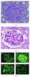
Figure 1
Figure 1
Two polar morphologic forms are representative of histological findings in lupus nephritis. (a) DPLN shows diffuse hypercellularity of endothelial cells and mesangial cells throughout the glomerulus. Peripheral capillary are thickened by subendothelial deposition of immune complex (x400, PAS staining). (b) On the other hand MLN shows generalized diffuse thickening of the peripheral capillary walls without prominent hypercellularity in the glomerulus (x400, PAS staining). (c) Glomerular IgG subclass deposits in a patient with MLN determined by immunofluorescence microscopy (x100). IgG subclass deposition was examined using FITC-conjugated mouse antihuman IgG subclass specific mAbs.
Figure 2
Figure 2
Representative two-color dot plots (anti-IL-4 PE versus anti-IFN-γ FITC) from analysis of intracellular cytokines by flow cytometry. Peripheral whole blood samples obtained from individuals were cultured for 4 hours with PMA and ionomycin in the presence of brefeldin A. The activated cells were fixed and stained with anti IFN-γ FITC, anti-IL-4 PE, and CD4-PerCP. The cells were then subjected to flow cytometric analysis on a FACScan flow cytometer (Becton Dickinson). Dot plots (a) and (b) are typical examples of FACS histograms which have demonstrated contrastive polarization to the Th1- and Th2-like cytokine responses, respectively. (a) histogram for one SLE patient (age/sex; 48/F) with DPLN. (b) histogram for another SLE patient (age/sex; 21/F) with MLN. The percentage of cells in each of the quadrants are shown in the right upper corners. The values of the Th1/Th2 ratio are shown in the top of each panel.
Figure 3
Figure 3
Distribution of the individual values of Th1/Th2 ratios in three groups control, DPLN, and MLN patients. Individual values of the ratio are plotted with the mean (bar) for each group, and (n) refers to the number of individuals in each group. The mean ± SD values were as follows. Healthy normal control 8.82 ± 4.45, DPLN 25.89 ± 18.98, and MLN 4.66 ± 4.64, respectively. The value of DPLN was significantly higher than those of normal and MLN. P values determined by Student's t-test.
Figure 4
Figure 4
MRL/lpr mice disrupted the WSX-1 gene developed disease essentially identical with human MLN. (a) A representative glomerulus of 36-week-old WSX-1−/− MRL/lpr mouse (x400, PAM staining). Arrows indicate spike formation. (b) Electron micrograph (x4000) of the glomerular capillary of the mouse shows numerous subepithelial electron-dense deposits in the basement membrane (arrows).

References

    1. Cochrane CG, Koffler D. Immune complex disease in experimental animals and man. Advances in Immunology. 1973;16(C):185–264. - PubMed
    1. Kotzin BL. Systemic lupus erythematosus. Cell. 1996;85(3):303–306. - PubMed
    1. Churg J, Bernstein J, Glassock RJ. Lupus nephritis. In: Churg J, Bernstein J, Glassock RJ, editors. Renal Disease: Classification and Atlas of Glomerular Disease. New York, NY, USA: Igaku-Shoin Medical; 1995. p. 151.
    1. Dixon FJ. The pathogenesis of glomerulonephritis. The American Journal of Medicine. 1968;44(4):493–498. - PubMed
    1. Weening JJ, D’Agati VD, Schwartz MM, et al. The classification of glomerulonephritis in systemic lupus erythematosus revisited. Kidney International. 2004;65(2):521–530. - PubMed

LinkOut - more resources