

Functional magnetic resonance in the evaluation of oesophageal motility disorders
- PMID: 21904543
- PMCID: PMC3166566
- DOI: 10.1155/2011/367639
Functional magnetic resonance in the evaluation of oesophageal motility disorders
Abstract
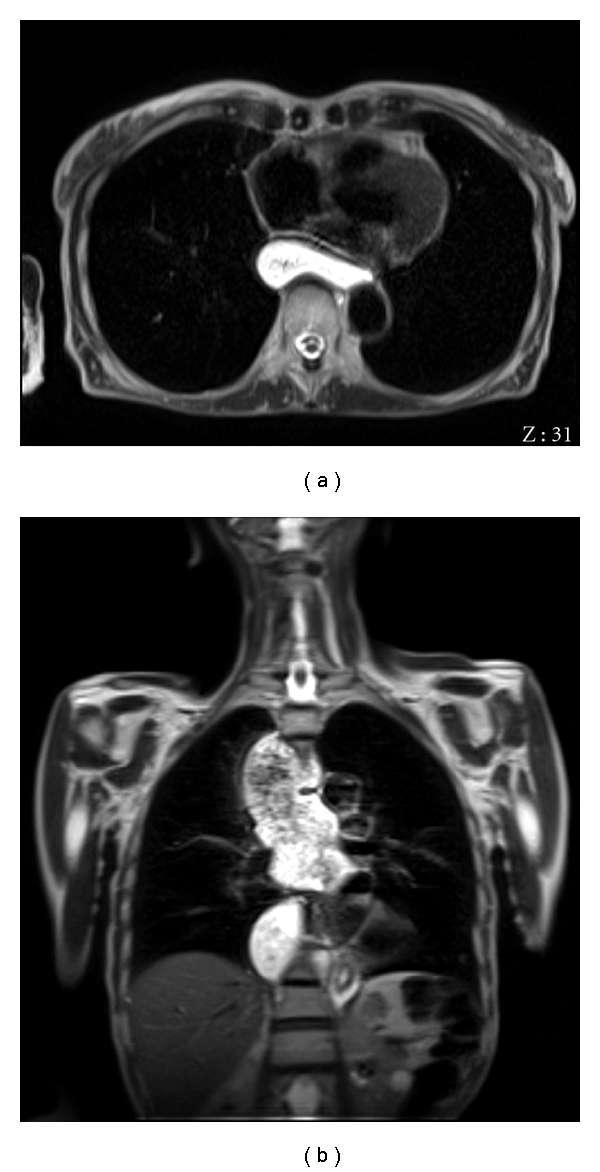
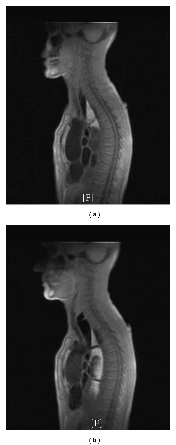
Functional magnetic resonance imaging (fMRI) has been recently proposed for the evaluation of the esophagus. Our aim is to assess the role of fMRI as a technique to assess morphological and functional parameters of the esophagus in patients with esophageal motor disorders and in healthy controls. Subsequently, we assessed the diagnostic efficiency of fMRI in comparison to videofluoroscopic and manometric findings in the investigation of patients with esophageal motor disorders. Considering that fMRI was shown to offer valuable information on bolus transit and on the caliber of the esophagus, variations of these two parameters in the different types of esophageal motor alterations have been assessed. fMRI, compared to manometry and videofluoroscopy, showed that a deranged or absent peristalsis is significantly associated with slower transit time and with increased esophageal diameter. Although further studies are needed, fMRI represents a promising noninvasive technique for the integrated functional and morphological evaluation of esophageal motility disorders.
Figures
References
-
- Richter JE. Oesophageal motility disorders. Lancet. 2001;358(9284):823–828. - PubMed
-
- Ghosh SK, Pandolfino JE, Zhang Q, Jarosz A, Shah N, Kahrilas PJ. Quantifying esophageal peristalsis with high-resolution manometry: a study of 75 asymptomatic volunteers. American Journal of Physiology. 2006;290(5):G988–G997. - PubMed
-
- Tutuian R, Castell DO. Combined multichannel intraluminal impedance and manometry clarifies esophageal function abnormalities: study in 350 patients. American Journal of Gastroenterology. 2004;99(6):1011–1019. - PubMed
-
- Prabhakar A, Levine MS, Rubesin S, Laufer I, Katzka D. Relationship between diffuse esophageal spasm and lower esophageal sphincter dysfunction on barium studies and manometry in 14 patients. American Journal of Roentgenology. 2004;183(2):409–413. - PubMed
-
- Fuller L, Huprich JE, Theisen J, et al. Abnormal esophageal body function: radiographic-manometric correlation. American Surgeon. 1999;65(10):911–914. - PubMed
LinkOut - more resources
Full Text Sources
