

White matter lesions defined by diffusion tensor imaging in older adults
- PMID: 21905080
- PMCID: PMC3177155
- DOI: 10.1002/ana.22484
White matter lesions defined by diffusion tensor imaging in older adults
Abstract
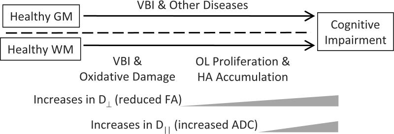
Objective: The cellular and molecular mechanisms underlying magnetic resonance imaging-defined white matter (WM) changes associated with age-related cognitive decline remain poorly defined. We tested the hypothesis that WM lesions in older adults, defined by diffusion tensor imaging (DTI), arise in the setting of vascular brain injury (VBI) and are characterized by increased free radical injury and aberrant oligodendrocyte lineage (OL) cell response to injury.
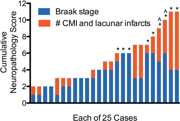
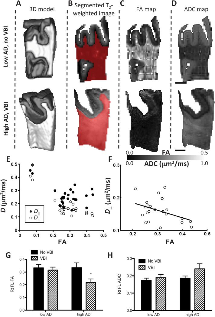
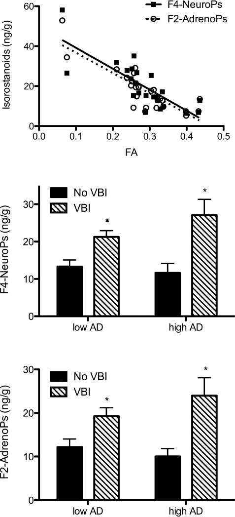
Methods: We undertook a multimodal analysis of prefrontal cortex (PFC) WM from 25 autopsies derived from a population-based cohort where VBI and Alzheimer disease (AD) frequently coincide. Ex vivo high field strength DTI measurements of fractional anisotropy (FA), apparent diffusion coefficient, and axial and radial (D(⊥) ) diffusivity were measured at high magnetic field strength (11.7T) and analyzed relative to quantitative in vivo biomarkers of free radical injury, an OL-specific marker Olig2, and histologic evaluation of hyaluronan (HA), an inhibitor of OL maturation.
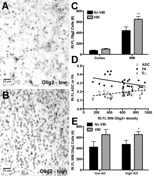
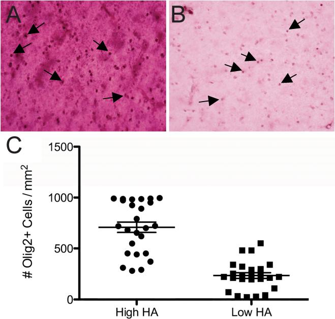
Results: Coincident AD and VBI showed significant association with lower FA and a robust relationship between decreasing FA and increasing D(⊥) . Free radical injury to docosahexaenoate and adrenate in PFC WM was significantly elevated in cases with VBI independent of AD, and was inversely correlated with FA. Similarly, increased density of Olig2-immunoreactive cells in PFC WM was significantly associated with VBI independent of AD and colocalized with regions enriched in HA.
Interpretation: DTI-defined PFC WM lesions in older individuals are characterized by free radical injury to myelin and neuroaxonal elements that coincides with pronounced expansion of the pool of OL cells in HA-rich regions.
Copyright © 2011 American Neurological Association.
Figures
Similar articles
-
Associations between white matter microstructure and amyloid burden in preclinical Alzheimer's disease: A multimodal imaging investigation.Neuroimage Clin. 2014 Feb 19;4:604-14. doi: 10.1016/j.nicl.2014.02.001. eCollection 2014. Neuroimage Clin. 2014. PMID: 24936411 Free PMC article.
-
Fractional anisotropy changes in Alzheimer's disease depend on the underlying fiber tract architecture: a multiparametric DTI study using joint independent component analysis.J Alzheimers Dis. 2014;41(1):69-83. doi: 10.3233/JAD-131829. J Alzheimers Dis. 2014. PMID: 24577476
-
Are functional deficits in concussed individuals consistent with white matter structural alterations: combined FMRI & DTI study.Exp Brain Res. 2010 Jul;204(1):57-70. doi: 10.1007/s00221-010-2294-3. Epub 2010 May 23. Exp Brain Res. 2010. PMID: 20496060 Free PMC article.
-
The role of diffusion tensor imaging and fractional anisotropy in the evaluation of patients with idiopathic normal pressure hydrocephalus: a literature review.Neurosurg Focus. 2016 Sep;41(3):E12. doi: 10.3171/2016.6.FOCUS16192. Neurosurg Focus. 2016. PMID: 27581308 Review.
-
The role of diffusion tensor imaging in the evaluation of ischemic brain injury - a review.NMR Biomed. 2002 Nov-Dec;15(7-8):561-9. doi: 10.1002/nbm.786. NMR Biomed. 2002. PMID: 12489102 Review.
Cited by
-
Digestion products of the PH20 hyaluronidase inhibit remyelination.Ann Neurol. 2013 Feb;73(2):266-80. doi: 10.1002/ana.23788. Epub 2013 Mar 5. Ann Neurol. 2013. PMID: 23463525 Free PMC article.
-
Astroglial NF-kB contributes to white matter damage and cognitive impairment in a mouse model of vascular dementia.Acta Neuropathol Commun. 2016 Aug 4;4(1):76. doi: 10.1186/s40478-016-0350-3. Acta Neuropathol Commun. 2016. PMID: 27487766 Free PMC article.
-
Hypertension: a harbinger of stroke and dementia.Hypertension. 2013 Nov;62(5):810-7. doi: 10.1161/HYPERTENSIONAHA.113.01063. Epub 2013 Aug 26. Hypertension. 2013. PMID: 23980072 Free PMC article. Review. No abstract available.
-
Unbiased Stereological Analysis of Reactive Astrogliosis to Estimate Age-Associated Cerebral White Matter Injury.J Neuropathol Exp Neurol. 2016 Jun;75(6):539-54. doi: 10.1093/jnen/nlw032. Epub 2016 May 3. J Neuropathol Exp Neurol. 2016. PMID: 27142644 Free PMC article.
-
Antioxidant treatment strategies for hyperphenylalaninemia.Metab Brain Dis. 2013 Dec;28(4):541-50. doi: 10.1007/s11011-013-9414-2. Epub 2013 May 9. Metab Brain Dis. 2013. PMID: 23657560 Review.
References
-
- Ylikoski A, Erkinjuntti T, Raininko R, Sarna S, Sulkava R, Tilvis R. White matter hyperintensities on MRI in the neurologically nondiseased elderly. Analysis of cohorts of consecutive subjects aged 55 to 85 years living at home. Stroke. 1995;26:1171–1177. - PubMed
-
- Breteler MM, van Swieten JC, Bots ML, Grobbee DE, Claus JJ, van den Hout JH, van Harskamp F, Tanghe HL, de Jong PT, van Gijn J, et al. Cerebral white matter lesions, vascular risk factors, and cognitive function in a population-based study: the Rotterdam Study. Neurology. 1994;44:1246–1252. - PubMed
-
- Longstreth WT, Jr., Dulberg C, Manolio TA, Lewis MR, Beauchamp NJ, Jr., O'Leary D, Carr J, Furberg CD. Incidence, manifestations, and predictors of brain infarcts defined by serial cranial magnetic resonance imaging in the elderly: the Cardiovascular Health Study. Stroke. 2002;33:2376–2382. - PubMed
-
- Gunning-Dixon FM, Raz N. The cognitive correlates of white matter abnormalities in normal aging: a quantitative review. Neuropsychology. 2000;14:224–232. - PubMed
-
- DeCarli C, Scheltens P. Structural brain imaging, in Vascular cognitive impairment. In: Erkinjuntti T, Gauthier S, editors. Vascular cognitive impairment. Martin Dunitz; London: 2002. pp. 433–457.
Publication types
MeSH terms
Substances
Grants and funding
- R01 NS054044/NS/NINDS NIH HHS/United States
- R01 AG031892/AG/NIA NIH HHS/United States
- R01 AG023801/AG/NIA NIH HHS/United States
- NS045737/NS/NINDS NIH HHS/United States
- K01 RR000163/RR/NCRR NIH HHS/United States
- RR000163/RR/NCRR NIH HHS/United States
- R01 NS045737/NS/NINDS NIH HHS/United States
- AG031892/AG/NIA NIH HHS/United States
- R37 NS045737/NS/NINDS NIH HHS/United States
- UL1 TR000128/TR/NCATS NIH HHS/United States
- P51 RR000163/RR/NCRR NIH HHS/United States
- U01 AG006781/AG/NIA NIH HHS/United States
- AG06781/AG/NIA NIH HHS/United States
LinkOut - more resources
Full Text Sources
Miscellaneous
