

Purification and characterization of hydroquinone dioxygenase from Sphingomonas sp. strain TTNP3
- PMID: 21906340
- PMCID: PMC3222310
- DOI: 10.1186/2191-0855-1-8
Purification and characterization of hydroquinone dioxygenase from Sphingomonas sp. strain TTNP3
Abstract
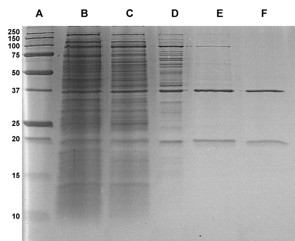
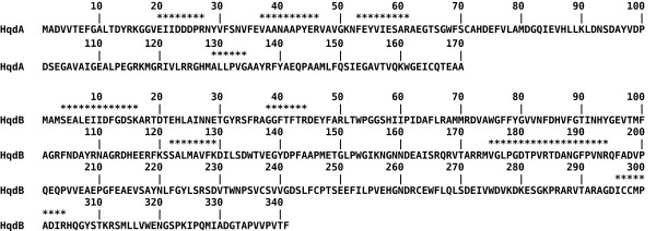
Hydroquinone-1,2-dioxygenase, an enzyme involved in the degradation of alkylphenols in Sphingomonas sp. strain TTNP3 was purified to apparent homogeneity. The extradiol dioxygenase catalyzed the ring fission of hydroquinone to 4-hydroxymuconic semialdehyde and the degradation of chlorinated and several alkylated hydroquinones. The activity of 1 mg of the purified enzyme with unsubstituted hydroquinone was 6.1 μmol per minute, the apparent Km 2.2 μM. ICP-MS analysis revealed an iron content of 1.4 moles per mole enzyme. The enzyme lost activity upon exposure to oxygen, but could be reactivated by Fe(II) in presence of ascorbate. SDS-PAGE analysis of the purified enzyme yielded two bands of an apparent size of 38 kDa and 19 kDa, respectively. Data from MALDI-TOF analyses of peptides of the respective bands matched with the deduced amino acid sequences of two neighboring open reading frames found in genomic DNA of Sphingomonas sp strain TTNP3. The deduced amino acid sequences showed 62% and 47% identity to the large and small subunit of hydroquinone dioxygenase from Pseudomonas fluorescens strain ACB, respectively. This heterotetrameric enzyme is the first of its kind found in a strain of the genus Sphingomonas sensu latu.
Figures
Similar articles
-
An unexpected gene cluster for downstream degradation of alkylphenols in Sphingomonas sp. strain TTNP3.Appl Microbiol Biotechnol. 2012 Feb;93(3):1315-24. doi: 10.1007/s00253-011-3451-8. Epub 2011 Jul 14. Appl Microbiol Biotechnol. 2012. PMID: 21755281
-
The crystal structures of native hydroquinone 1,2-dioxygenase from Sphingomonas sp. TTNP3 and of substrate and inhibitor complexes.Biochim Biophys Acta Proteins Proteom. 2017 May;1865(5):520-530. doi: 10.1016/j.bbapap.2017.02.013. Epub 2017 Feb 20. Biochim Biophys Acta Proteins Proteom. 2017. PMID: 28232026
-
Crystallization and preliminary X-ray crystallographic analysis of hydroquinone dioxygenase from Sphingomonas sp. TTNP3.Acta Crystallogr Sect F Struct Biol Cryst Commun. 2012 May 1;68(Pt 5):588-90. doi: 10.1107/S1744309112012341. Epub 2012 Apr 21. Acta Crystallogr Sect F Struct Biol Cryst Commun. 2012. PMID: 22691794 Free PMC article.
-
Hydroquinone dioxygenase from pseudomonas fluorescens ACB: a novel member of the family of nonheme-iron(II)-dependent dioxygenases.J Bacteriol. 2008 Aug;190(15):5199-209. doi: 10.1128/JB.01945-07. Epub 2008 May 23. J Bacteriol. 2008. PMID: 18502867 Free PMC article.
-
The degradation of alkylphenols by Sphingomonas sp. strain TTNP3 - a review on seven years of research.N Biotechnol. 2012 Nov 15;30(1):88-95. doi: 10.1016/j.nbt.2012.07.008. Epub 2012 Jul 27. N Biotechnol. 2012. PMID: 22842087 Review.
Cited by
-
High activity catechol 1,2-dioxygenase from Stenotrophomonas maltophilia strain KB2 as a useful tool in cis,cis-muconic acid production.Antonie Van Leeuwenhoek. 2013 Jun;103(6):1297-307. doi: 10.1007/s10482-013-9910-8. Epub 2013 Mar 28. Antonie Van Leeuwenhoek. 2013. PMID: 23536173 Free PMC article.
-
Efficient degradation of hydroquinone by a metabolically engineered Pseudarthrobacter sulfonivorans strain.Arch Microbiol. 2022 Sep 1;204(9):588. doi: 10.1007/s00203-022-03214-z. Arch Microbiol. 2022. PMID: 36048304
-
Branching of the p-nitrophenol (PNP) degradation pathway in burkholderia sp. Strain SJ98: Evidences from genetic characterization of PNP gene cluster.AMB Express. 2012 Jun 8;2(1):30. doi: 10.1186/2191-0855-2-30. AMB Express. 2012. PMID: 22681853 Free PMC article.
-
Metabolism of 2-chloro-4-nitrophenol in a gram negative bacterium, Burkholderia sp. RKJ 800.PLoS One. 2012;7(6):e38676. doi: 10.1371/journal.pone.0038676. Epub 2012 Jun 6. PLoS One. 2012. PMID: 22701692 Free PMC article.
-
Hydroquinone: environmental pollution, toxicity, and microbial answers.Biomed Res Int. 2013;2013:542168. doi: 10.1155/2013/542168. Epub 2013 Jul 15. Biomed Res Int. 2013. PMID: 23936816 Free PMC article. Review.
References
LinkOut - more resources
Full Text Sources
Molecular Biology Databases
