Review
. 2011:99:23-50.
doi: 10.1016/B978-0-12-387003-2.00002-1.

Homeostatic control of neural activity: a Drosophila model for drug tolerance and dependence

Affiliations
Review

Homeostatic control of neural activity: a Drosophila model for drug tolerance and dependence

Alfredo Ghezzi et al. Int Rev Neurobiol. 2011.

Abstract

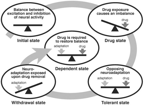
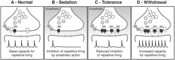
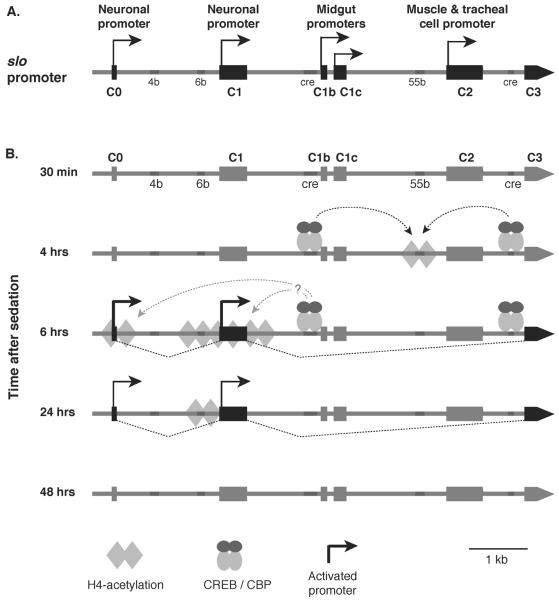
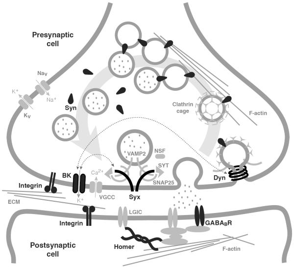
Drug addiction is a complex condition of compulsive drug use that results in devastating physical and social consequences. Drosophila melanogaster has recently emerged as a valuable genetic model for investigating the mechanisms of addiction. Drug tolerance is a measurable endophenotype of addiction that can be easily generated and detected in animal models. The counteradaptive theory for drug dependence postulates that the homeostatic adaptations that produce drug tolerance become counteradaptive after drug clearance, resulting in symptoms of dependence. In flies, a single sedation with ethanol or with an organic solvent anesthetic (benzyl alcohol) induces functional tolerance, an adaptation of the nervous system that reduces the effect of these neural depressants. Here we review the role of the BK channel gene (slo) and genes that encode other synaptic proteins in the process of producing functional tolerance. These proteins are predicted to be part of an orchestrated response that involves specific interactions across a highly complex synaptic protein network. The response of the slo gene to drug exposure and the consequence of induced slo expression fit nicely the tenets of the counteradaptive theory for drug tolerance and dependence. Induction of slo expression represents an adaptive process that generates tolerance because it enhances neuronal excitability, which counters the sedative effects of the drugs. After drug clearance, however, the increase in slo expression leads to an allostatic withdrawal state that is characterized by an increase in the susceptibility for seizure. Together, these results demonstrate a common origin for development of drug tolerance and withdrawal hyperexcitability in Drosophila.


Figures

Figure 1
Figure 1. Homeostatic counter-adaptive model of drug tolerance and dependence
In the counter-adaptive model of drug tolerance and dependence, the carefully controlled balance between excitation and inhibition of neural activity in the brain (Initial state) is significantly altered by exposure to a psychoactive drug, creating a state of activity imbalance (Drug state). In an attempt to restore the balance of neural activity, homeostatic neuroadaptive mechanisms are activated (Tolerant state). After drug clearance, the homeostatic neuroadaptation is exposed, resulting in an opposing unbalanced state (Withdrawal state), leading back to the requirement of the drug to restore balance (Dependent state). Continuing use of the drug leads to further adaptation and an intensified requirement for the drug, resulting in a spiraling negative feed-forward cascade. Figure adapted from Littleton (1998).
Figure 2
Figure 2. Benzyl alcohol tolerance assay
Shown are sequential pictures of two vials of flies taken at different time points of sedation and recovery from 0.4 % benzyl alcohol exposure. The vial on the left in every picture contains naïve flies, which have never been treated before; this is their first exposure (1st). The vial on the right contains flies that were previously sedated (24 hours earlier) with a similar dose of benzyl alcohol; this is their second exposure (2nd). The time point at which each picture was taken is indicated under each picture in minutes after start of the treatment. Time points from 1 to 9 minutes (white text over black) are in the presence of the solvent. The solvent has been removed at 10 minutes. Time points from 10 to 30 minutes (black text over white) are during the recovery in a solvent-free tube. Although both groups knock down simultaneously, flies recovering from the 2nd sedation recover negative geotaxis at earlier time points than flies recovering from their 1st sedation do.
Figure 3
Figure 3. Quantification of benzyl alcohol tolerance
Shown are knock-down and recovery curves of wild-type flies after one (1st exposure) or two (2nd exposure) exposures to 0.4% benzyl alcohol. Values are plotted as percentage of flies climbing during sedation with benzyl alcohol (gray background) and during recovery from sedation (white background). Time between exposures is 24 hours. Significant difference is determined by the log-rank test (Error bars are SEM for each data point).
Figure 4
Figure 4. Model for how homeostatic regulation of BK channels contributes to both drug tolerance and withdrawal
A) In a naïve, no drug state, neurons of the giant fiber pathway in Drosophila exhibit a basal capacity for repetitive firing when evoked by high frequency stimulation (Normal). B) The capacity for repetitive firing is significantly inhibited by exposure to sedative drugs (Sedation), leading to the overall depression of neural activity characteristic of sedation. C) Drug exposure induces expression of presynaptic BK channels as part of a homeostatic response to sedation. Increased BK channel activity enhances the capacity for repetitive firing, leading to a reduced effect of the anesthetic on neural firing during a subsequent exposure (Tolerance). D) After drug clearance, however, another effect of increased BK channel expression is unmasked, resulting in an enhanced neural excitability in the form of increased basal firing capacity and an increased susceptibility for seizures (Withdrawal). The electrophysiological traces shown here are schematic representations of hypothetical data, not real traces.
Figure 5
Figure 5. Drug-induced chromatin remodeling at the slo transcriptional control region
A) Shown is the 7 kb transcriptional control region of the slo gene. This gene has at least five alternative tissue-specific promoters: two neuronal promoters (C0 and C1), two midgut promoters (C1b and C1c), and one muscle cell and tracheal cell specific promoter (C2). In addition, this region contains several DNA elements that are highly conserved across different Drosophila species (4b, 6b, 55b) and two CREB response elements (cre). B) Dynamic histone H4 acetylation changes across the slo transcriptional control region after benzyl alcohol sedation. No change in acetylation is detected 30 minutes after sedation. At 4 hours, an increase in acetylation is detected with a peak centered over the 55b element. This event is dependent on binding of phospho-CREB at the two CRE sites. After six hours, the acetylation peak relocates to the neural promoter area with a small peak around C0 and a broad peak around C1 and mRNA expression from these promoters become evident. After 24 hours, the histone acetylation peak becomes focused at the 6b element, and mRNA expression decays. By 48 hours, histone acetylation and mRNA expression return to non-sedated control level.
Figure 6
Figure 6. Synaptic model of drug tolerance
Shown is a schematic representation of a synapse current knowledge of putative interactions between synaptic proteins. Synaptic proteins that have been implicated in the development of tolerance to alcohol and anesthetics are displayed and labeled in black. These proteins include Synapsins (Syn), Dynamin (Dyn), Homer, Integrins, BK channels (BK), Syntaxin 1A (Syx), and the GABAB receptor (GABABR). Proteins and structures in gray are included to provide context. Thin dotted lines with arrows denote known protein-protein interactions. Figure adapted from Gorini et al. (2010).

Similar articles

Cited by

References

    1. Al-Hasan YM, Krishnan HR, Ghezzi A, Prado FJ, 3rd., Robles RB, Atkinson NS. Tolerance to Anesthesia Depends on Synaptic Proteins. Behav. Genet. 2011 DOI 10.1007/s10519-011-9451-8. - PMC - PubMed
    1. Allada R, Nash HA. Drosophila melanogaster as a model for study of general anesthesia: the quantitative response to clinical anesthetics and alkanes. Anesth. Analg. 1993;77:19–26. - PubMed
    1. Allis CD, Jenuwein T, Reinberg D, Caparros M-L. Epigenetics. Cold Spring Harbor Laboratory Press; Woodbury, NY: 2007.
    1. Ankney PF. A note on barometric pressure and behavior in Drosophila pseudoobscura. Behav. Genet. 1984;14:315–317. - PubMed
    1. Atkinson NS. Tolerance in Drosophila. J. Neurogenet. 2009;23:293–302. - PMC - PubMed

Publication types

MeSH terms

Substances