. 2011 Dec;232(2):176-84.
doi: 10.1016/j.expneurol.2011.08.021. Epub 2011 Aug 30.

Altered position of cell bodies and fibers in the ventromedial region in SF-1 knockout mice

Affiliations

Altered position of cell bodies and fibers in the ventromedial region in SF-1 knockout mice

Tomaz Büdefeld et al. Exp Neurol. 2011 Dec.

Abstract

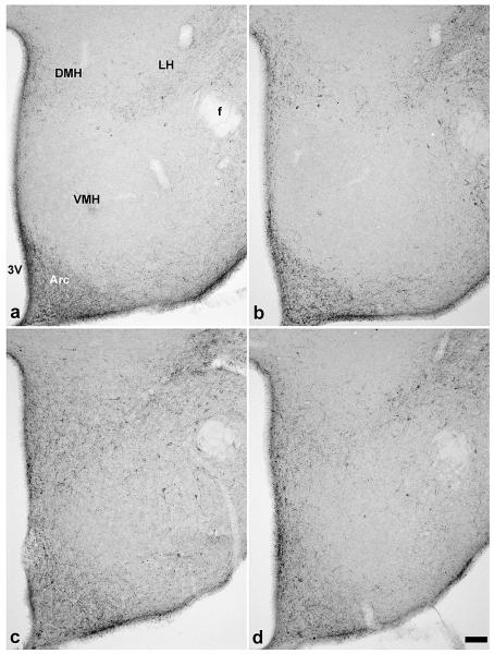
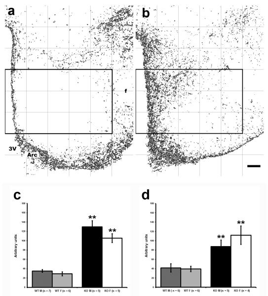
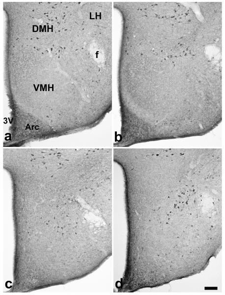
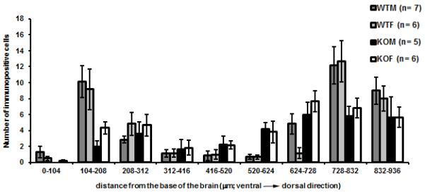
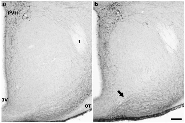
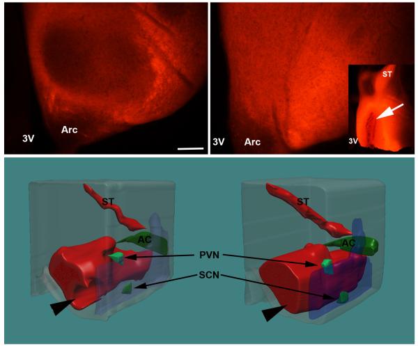
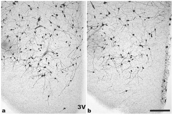
The ventromedial nucleus of the hypothalamus (VMH) is a key cell group in the medial-basal hypothalamus that participates in the regulation of energy balance. Previous studies have shown that the cellular organization of the VMH is altered in mice with a disruption of the steroidogenic factor-1 (NR5a1) gene (SF-1 KO mice). The present study examined orexigenic/anorexigenic peptides (neuropeptide Y (NPY), agouti-related peptide (AgRP) and cocaine- and amphetamine-regulated transcript (CART)) and neural connections to and from the VMH in SF1 KO mice. NeuroVue tracing and Golgi staining were used to evaluate connections between the preoptic area (POA) and VMH and the orientation of dendrites in the VMH, respectively. Results of this study reveal changes in the cytoarchitecture of the region of the VMH with respect to the distribution of immunoreactive NPY, AgRP and CART. In WT mice projections from the POA normally surround the VMH while in SF-1 KO mice, projections from the POA stream through the region that would otherwise be VMH. Golgi impregnation of the region revealed fewer dendrites with ventrolateral orientations and in general, more variable dendritic orientations in SF-1 KO mice providing additional evidence that the connectivity of cells in the region is likely altered due to the cellular rearrangements consequent to disruption of the NR5a1 gene. In conclusion, this study greatly extends the data showing that the morphology of the regions containing the VMH is disrupted in SF-1 KO mice and suggests that changes in the location of cells or fibers containing NPY, AgRP and CART may, in part, account for changes in body weight homeostasis in these mice.


Figures

Figure 1
Figure 1
Digital images show NPY immunoreactivity in the medial hypothalamus in sections 1.7 mm caudal to bregma was limited to the arcuate (Arc) and dorsomedial nuclei (DMH) in WT male (a) and female (b) mice. In contrast, in SF-1 KO mice (male; c and female; d), NPY immunoreactivity was detected in the area of ventromedial nucleus (VMH), which was devoid of these fibers in WT mice of both sexes. 3V- the third ventricle, Arc– arcuate nucleus, f- fornix, LH- lateral hypothalamus, DMH– dorsomedial nucleus of the hypothalamus; scale bar in d represents 100 μm for all panels.
Figure 2
Figure 2
Digital images showing AgRP- ir fibers in WT male mouse (a) and in corresponding sections in SF-1 KO male mouse (b) in the box 5×3 covering VMH nucleus as shown after processing for intensity thresholding (a, b). Graphs (c, d) show the quantification of AgRP and NPY- ir area (mean ± S.E.M.) that indicate significantly greater immunoreactive area in the designated VMH region in SF-1 KO mice (c- AgRP, p < 0.001; **) and (d- NPY, p < 0.001; **). 3V- the third ventricle, Arc- arcuate nucleus, f- fornix, scale bar in b represents 100μm for both panels a and b.
Figure 3
Figure 3
Digital images show CART- ir cells in the medial hypothalamus in sections 1.7 mm caudal to bregma in WT male (a), WT female (b), SF-1 KO male (c) and SF-1 KO female (d) mice. Location of CART- ir cells is altered in SF-1 KO mice, where CART -ir cells are shifted ventromedially into the corresponding dorsomedial part of VMH area. Number of CART- ir cells did not differ between all four groups. 3V- the third ventricle, Arc- arcuate ucleus, DMH- dorsomedial nucleus of the hypothalamus, f- fornix, LH- lateral hypothalamus, VMH- ventromedial hypothalamus, bar: 100 μm 4
Figure 4
Figure 4
The graph shows the number (mean ± S.E.M.) of CART- ir cells in VMH region in WT mice and SF-1 KO mice as a function of distance from the base of the brain. Three-way ANOVA (age x sex) with the distance from the base of the brain (in 104μm bands or rows) as repetitive factor showed statistically significant differences between genotypes (* p< 0.001).
Figure 5
Figure 5
Digital images show AVP immunoreactivity in the medial basal hypothalamus in the coronal sections approximately 0.9 mm caudal to bregma in representative figures for each genotype (WT - a, and SF-1 KO - b). The position of immunoreactive fibers is altered in the ventrolateral part of the region in SF-1 KO (b) in comparison to WT (a) mice regardless of sex. AVP- ir fibers that project toward the median eminence are positioned more medially toward the third ventricle and more perpendicularly to the base of a brain in SF-1 KO mice (arrow). 3V- the third ventricle, f- fornix, OT- optic tract, PVH- paraventricular nucleus of hypothalamus, scale bar in b represents 100μm for both a and b.
Figure 6
Figure 6
Digital images show representative coronal brain sections through the VMH area with NeuroVue labeled fibers originating in the POA. In WT mice (a) central part of the VMH nucleus remained unlabeled (seen as darker oval shape) while in SF-1 KO mice (b) labeled neuronal fibers from POA filled the entire the region. This is further illustrated by 3D reconstruction of serial sections from POA to the caudal VMH region (c and d); 3V- the third entricle, Arc- arcuate nucleus, PVH- 1 paraventricular nucleus, SCN– suprachiasmatic nucleus, AC– anterior commissure, ST– stria terminalis; scale bar: 100μm).
Figure 7
Figure 7
Digital images show representative coronal brain sections through the VMH area with Golgi impregnated neurons. In WT mice (a) most primary dendrites in the ventrolateral part of the VMH were oriented ventrolaterally while in the VMH from SF-1 KO mice (b), primary dendrites were oriented in numerous directions (3V- the third ventricle, bar: 100 μm).

Similar articles

Cited by

References

    1. Brobeck JR. Mechanism of the development of obesity in animals with hypothalamic lesions. Physiol. Rev. 1946;26:541–559. - PubMed
    1. Broberger C. Hypothalamic cocaine- and amphetamine-regulated transcript (CART) neurons: histochemical relationship to thyrotropin-releasing hormone, melanin concentrating hormone, orexin/hypocretin and neuropeptide Y. Brain Res. 1999;848:101–113. - PubMed
    1. Broberger C, Johansen J, Johansson C, Schalling M, Hokfelt T. The neuropeptide Y/agouti gene-related protein (AGRP) brain circuitry in normal, anorectic, and monosodium glutamate-treated mice. Proc. Natl. Acad. Sci. U. S. A. 1998;95:15043–15048. - PMC - PubMed
    1. Broberger C, Visser TJ, Kuhar MJ, Hokfelt T. Neuropeptide Y innervation and neuropeptide-Y-Y1-receptor-expressing neurons in the paraventricular hypothalamic nucleus of the mouse. Neuroendocrinology. 1999;70:295–305. - PubMed
    1. Budefeld T, Grgurevic N, Tobet SA, Majdic G. Sex differences in brain developing in the presence or absence of gonads. Dev Neurobiol. 2008;68:981–995. - PMC - PubMed

Publication types

MeSH terms

Substances