

Neural correlates of blink suppression and the buildup of a natural bodily urge
- PMID: 21906689
- PMCID: PMC3230735
- DOI: 10.1016/j.neuroimage.2011.08.050
Neural correlates of blink suppression and the buildup of a natural bodily urge
Abstract
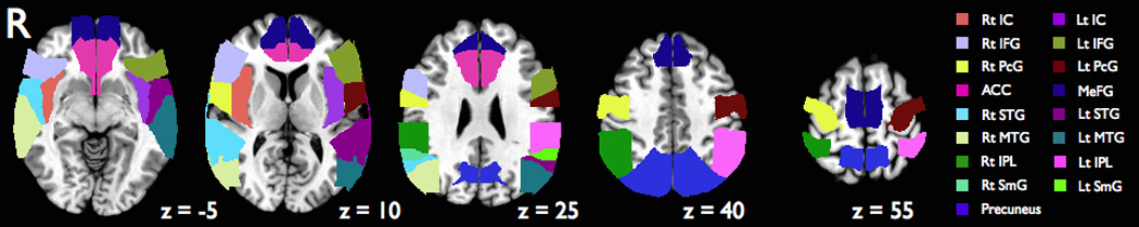
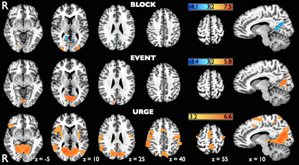
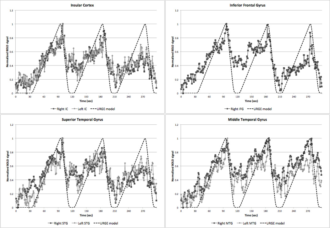
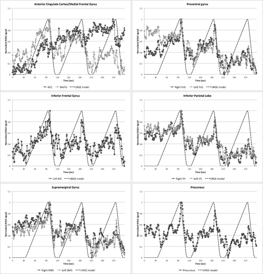
Neuroimaging studies have elucidated some of the underlying physiology of spontaneous and voluntary eye blinking; however, the neural networks involved in eye blink suppression remain poorly understood. Here we investigated blink suppression by analyzing fMRI data in a block design and event-related manner, and employed a novel hypothetical time-varying neural response model to detect brain activations associated with the buildup of urge. Blinks were found to activate visual cortices while our block design analysis revealed activations limited to the middle occipital gyri and deactivations in medial occipital, posterior cingulate and precuneus areas. Our model for urge, however, revealed a widespread network of activations including right greater than left insular cortex, right ventrolateral prefrontal cortex, middle cingulate cortex, and bilateral temporo-parietal cortices, primary and secondary face motor regions, and visual cortices. Subsequent inspection of BOLD time-series in an extensive ROI analysis showed that activity in the bilateral insular cortex, right ventrolateral prefrontal cortex, and bilateral STG and MTG showed strong correlations with our hypothetical model for urge suggesting these areas play a prominent role in the buildup of urge. The involvement of the insular cortex in particular, along with its function in interoceptive processing, helps support a key role for this structure in the buildup of urge during blink suppression. The right ventrolateral prefrontal cortex findings in conjunction with its known involvement in inhibitory control suggest a role for this structure in maintaining volitional suppression of an increasing sense of urge. The consistency of our urge model findings with prior studies investigating the suppression of blinking and other bodily urges, thoughts, and behaviors suggests that a similar investigative approach may have utility in fMRI studies of disorders associated with abnormal urge suppression such as Tourette syndrome and obsessive-compulsive disorder.
Copyright © 2011 Elsevier Inc. All rights reserved.
Figures
References
-
- Aron AR, Robbins TW, Poldrack RA. Inhibition and the right inferior frontal cortex. Trends in Cognitive Sciences. 2004;8(4):170–177. - PubMed
-
- Astafiev SV, Stanley CM, Shulman GL, Corbetta M. Extrastriate body area in human occipital cortex responds to the performance of motor actions. Nature Neuroscience. 2004;7(5):542–548. - PubMed
-
- Banzett RB, Mulnier HE, Murphy K, Rosen SD, Wise RJ, Adams L. Breathlessness in humans activates insular cortex. Neuroreport. 2000;11(10):2117–2120. - PubMed
-
- Bentivoglio AR, Bressman SB, Cassetta E, Carretta D, Tonali P, Albanese A. Analysis of blink rate patterns in normal subjects. Movement Disorders. 1997;12(6):1028–1034. - PubMed
-
- Bernal B, Altman N. Neural networks of motor and cognitive inhibition are dissociated between brain hemispheres: An fMRI study. International Journal of Neuroscience. 2009;119(10):1848–1880. - PubMed
Publication types
MeSH terms
Grants and funding
LinkOut - more resources
Full Text Sources
Medical
