. 2011 Dec;32(35):9180-7.
doi: 10.1016/j.biomaterials.2011.08.050. Epub 2011 Sep 8.

Pluripotent stem cell-derived cardiac tissue patch with advanced structure and function

Affiliations

Pluripotent stem cell-derived cardiac tissue patch with advanced structure and function

Brian Liau et al. Biomaterials. 2011 Dec.

Abstract

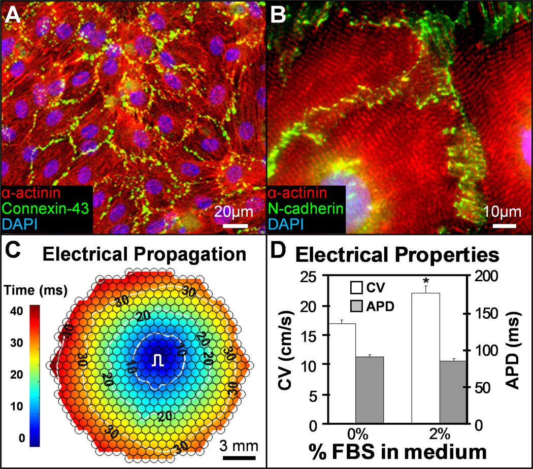
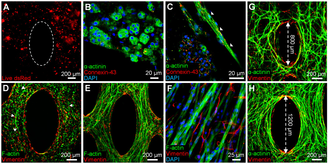
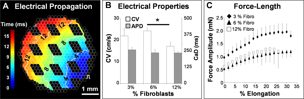
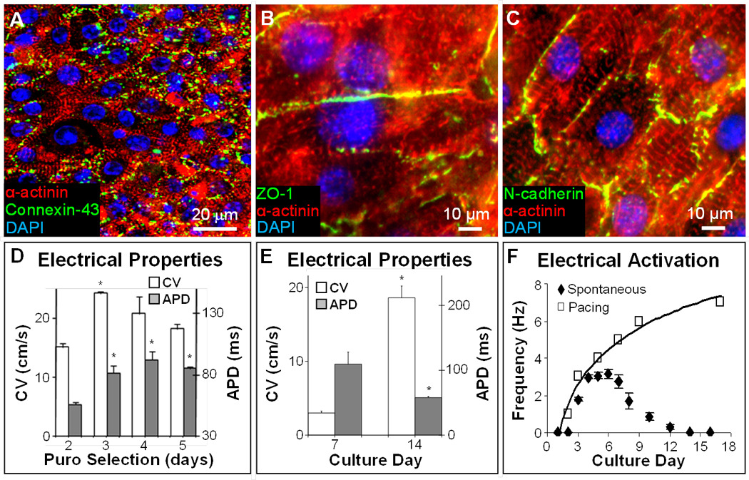
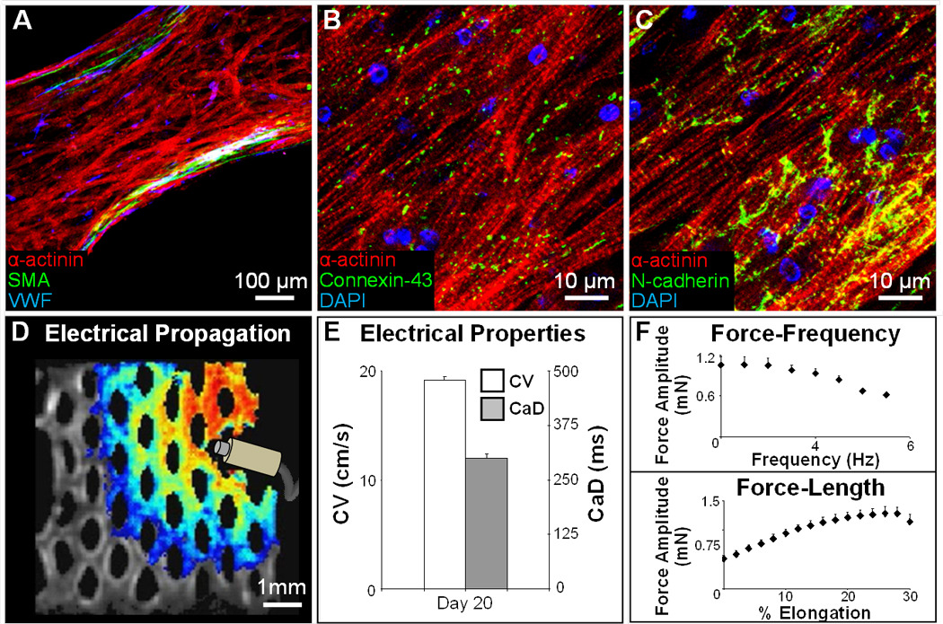
Recent advances in pluripotent stem cell research have provided investigators with potent sources of cardiogenic cells. However, tissue engineering methodologies to assemble cardiac progenitors into aligned, 3-dimensional (3D) myocardial tissues capable of physiologically relevant electrical conduction and force generation are lacking. In this study, we introduced 3D cell alignment cues in a fibrin-based hydrogel matrix to engineer highly functional cardiac tissues from genetically purified mouse embryonic stem cell-derived cardiomyocytes (CMs) and cardiovascular progenitors (CVPs). Procedures for CM and CVP derivation, purification, and functional differentiation in monolayer cultures were first optimized to yield robust intercellular coupling and maximize velocity of action potential propagation. A versatile soft-lithography technique was then applied to reproducibly fabricate engineered cardiac tissues with controllable size and 3D architecture. While purified CMs assembled into a functional 3D syncytium only when supplemented with supporting non-myocytes, purified CVPs differentiated into cardiomyocytes, smooth muscle, and endothelial cells, and autonomously supported the formation of functional cardiac tissues. After a total culture time similar to period of mouse embryonic development (21 days), the engineered cardiac tissues exhibited unprecedented levels of 3D organization and functional differentiation characteristic of native neonatal myocardium, including: 1) dense, uniformly aligned, highly differentiated and electromechanically coupled cardiomyocytes, 2) rapid action potential conduction with velocities between 22 and 25 cm/s, and 3) significant contractile forces of up to 2 mN. These results represent an important advancement in stem cell-based cardiac tissue engineering and provide the foundation for exploiting the exciting progress in pluripotent stem cell research in the future tissue engineering therapies for heart disease.


Figures

Figure 1
Figure 1
mESC-CMs form highly functional 2D monolayers. A–B, Under optimized conditions, pure mESC-CMs in 1–2 week old confluent monolayers exhibit well-defined sarcomeric structure and robust intercellular coupling via electrical (A) and mechanical (B) junctions. C, Point pacing (pulse sign) in mESC-CM monolayers elicits rapid and uniform action potential spread (see also Supplementary movie 2). Small circles denote 504 recording sites. Isochrones of cell activation (white circles) are labeled in milliseconds. d. Addition of 2% serum in culture media improves conduction velocity (CV) without affecting action potential duration (APD). *P < 0.05, one-tailed Student’s t-test; n = 6.
Figure 2
Figure 2
mESC-CMs in 3D cardiac patches spread and interconnect only in the presence of fibroblasts. A, Pure mESC-CMs in 14 day old tissue patches appear round and clustered. Dotted ellipse delineates the pore boundary created by the PDMS mold. B, mESC-CM clusters contain rounded, cross-striated cardiomyocytes (green) interconnected with connexin-43 gap junctions (red). C, Aligned cells are occasionally found only at the gel boundary (white arrowheads). D, In the presence of neonatal rat ventricular fibroblasts (red), mESC-CMs (green) start spreading (white arrowheads) by culture day 7. E, At day 14, mESC-CMs in co-cultured patches are extensively aligned. F, Fibroblasts around formed cardiac bundles show no preferential alignment. G,H, Degree of local cell alignment can be controlled by varying the length of microfabricated posts.
Figure 3
Figure 3
Co-cultured mESC-CM+fibroblast patches exhibit advanced electrical and mechanical function. A, Optical mapping revealed that co-cultured tissue patches supported fast and uniform action potential propagation (see also Supplementary movie 4). The void spaces in the isochrone map correspond to tissue pores. B, Conduction velocity (CV) and calcium transient duration (CaD) as a function of the initial fraction of fibroblasts (e.g., CV = 241 ± 16 mm/s for 6% fibroblasts). CV of 6% and 12% fibroblast conditions differed significantly. *P < 0.05, one-tailed Student’s t-test assuming unequal variances, n = 3. C, Co-culture tissue patches exhibited rising force-length relationships reminiscent of the Frank-Starling law in whole hearts. Percent elongation is shown relative to tissue length during culture (7 mm).
Figure 4
Figure 4
mESC-CVP monolayers exhibit robust electromechanical coupling and fast electrical propagation. A–C, After 7–14 days of culture, confluent mESC-CVP monolayers contain cross-striated cardiomyocytes that robustly express connexin-43 (A), zona occludens 1 (B), and N-cadherin (C). D, Electrophysiological parameters in 14-day old monolayers made using mESC-CVPs purified by selection with 5µg/ml puromycin (puro). The three-day puromycin selection yielded optimum conduction velocities (CV = 242 ± 4 mm/s) and was used in all further studies. *P < 0.05 relative to corresponding parameters for 2-day puromycin selection; ANOVA with Student-Newman-Keuls, n = 3–6. E–F, Functional cardiac differentiation of mESC-CVPs with time in culture was associated with increased CV and decreased APD (E), as well as decreased spontaneous beating rate (closed diamonds) and increased maximum rate of electrical pacing (open squares) yielding steady 1:1 capture (F). *P < 0.05 relative to day 7; one-tailed Student’s t-test, n = 3–5.
Figure 5
Figure 5
mESC-CVPs can autonomously form a functional 3D cardiac patch. A, Three-week old tissue patch with aligned cardiomyocytes (red), and dispersed endothelial (blue) and smooth muscle (green) cells. SMA; smooth muscle actin. VWF; Von Willebrand factor. B–C, mESC-CVP derived cardiomyocytes in tissue patches are cross-striated and robustly coupled via electrical (B) and mechanical (C) junctions. D, A snapshot of a uniformly propagating action potential (see Supplementary movie 5) induced by a point electrode. E–F, After 3 weeks in culture, mESC-CVP patches exhibit high CVs (E), significant contractile forces, and physiological force-frequency and force-length relationships (F). Mean ± s.e.m; n=3.

Similar articles

Cited by

References

    1. Pfeffer MA, Braunwald E. Ventricular remodeling after myocardial infarction. Experimental observations and clinical implications. Circulation. 1990;81:1161–1172. - PubMed
    1. Langer R, Vacanti JP. Tissue engineering. Science. 1993;260:920–926. - PubMed
    1. Zimmermann WH, Melnychenko I, Wasmeier G, Didie M, Naito H, Nixdorff U, et al. Engineered heart tissue grafts improve systolic and diastolic function in infarcted rat hearts. Nat Med. 2006;12:452–458. - PubMed
    1. Chien KR, Domian IJ, Parker KK. Cardiogenesis and the complex biology of regenerative cardiovascular medicine. Science (New York, NY. 2008;322:1494–1497. - PubMed
    1. Laflamme MA, Chen KY, Naumova AV, Muskheli V, Fugate JA, Dupras SK, et al. Cardiomyocytes derived from human embryonic stem cells in pro-survival factors enhance function of infarcted rat hearts. Nat Biotechnol. 2007;25:1015–1024. - PubMed

Publication types