

Organization of the smallest eukaryotic spindle
- PMID: 21906950
- PMCID: PMC3234289
- DOI: 10.1016/j.cub.2011.08.021
Organization of the smallest eukaryotic spindle
Abstract
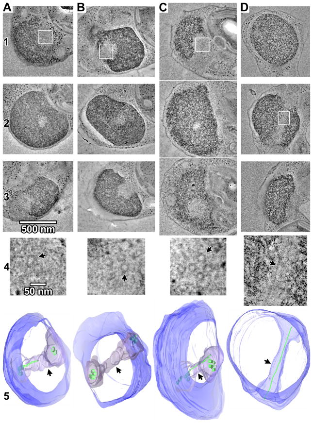
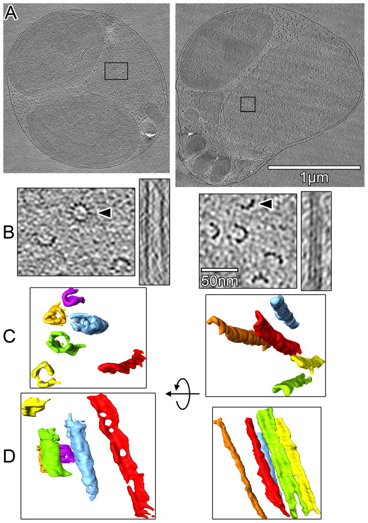
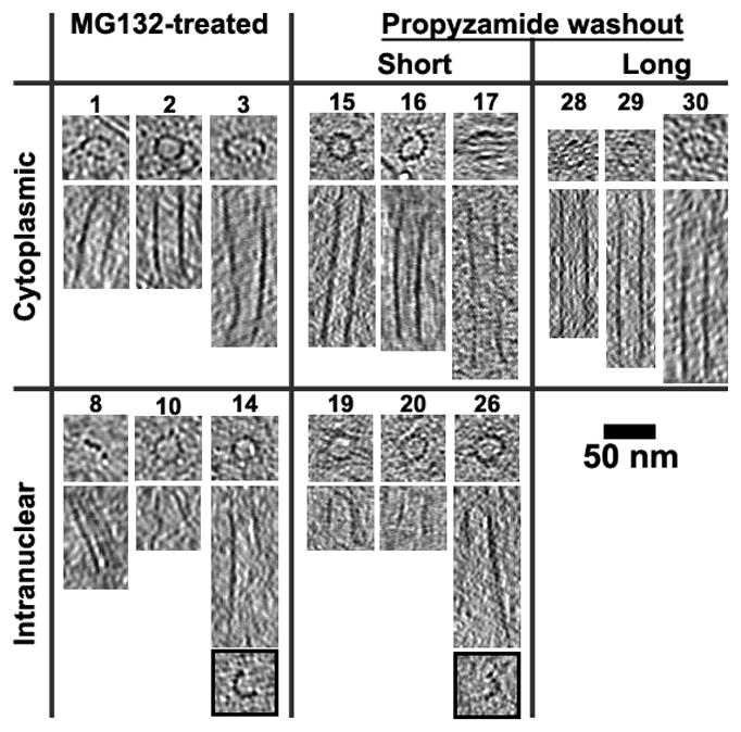
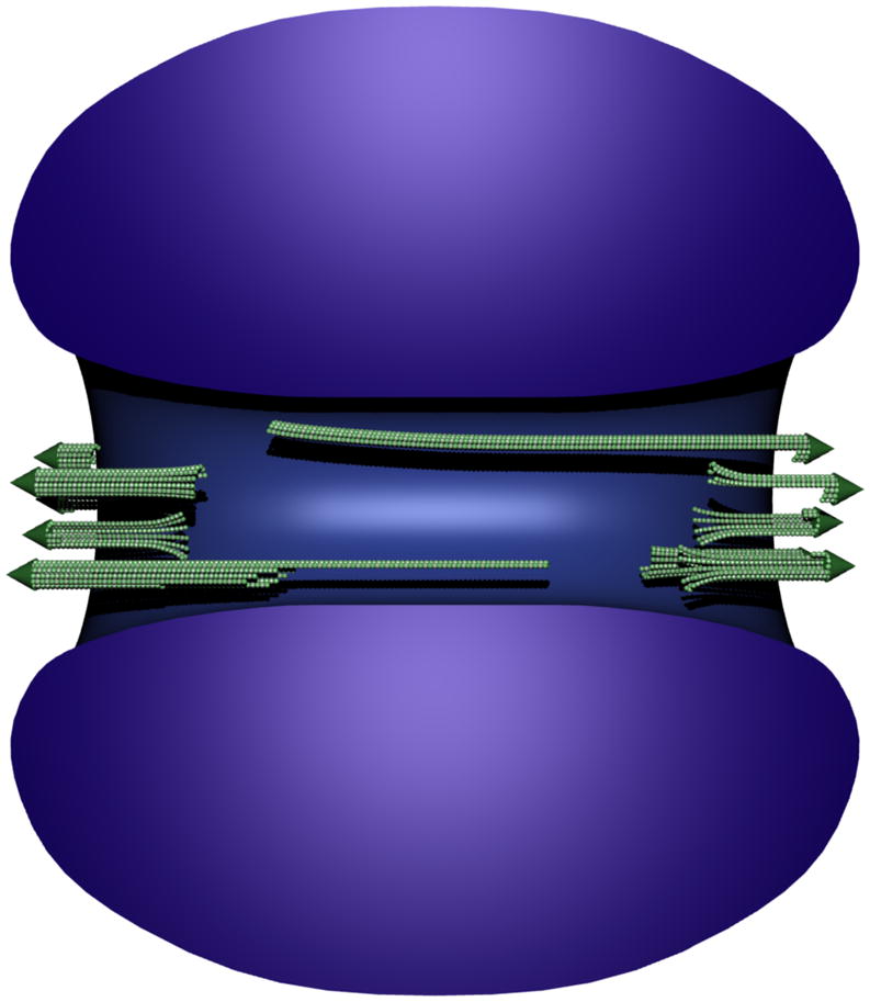
In metazoans, plants, and fungi, the spindle checkpoint delays mitosis until each chromosome is attached to one or more of its own kinetochore microtubules (kMTs). Some unicellular eukaryotes, however, have been reported to have fewer kMTs than chromosomes [1-5]. If this is the case, it is unclear how the spindle checkpoint could be satisfied. In the vast majority of the previous studies, mitotic cells were chemically fixed at room temperature, but this does not always preserve dynamic and/or small structures like spindle MTs and kinetochores [6]. Indeed, later higher-resolution studies have reversed some earlier claims [7-11]. Here we show that in Ostreococcus tauri (the smallest eukaryote known), mitosis does involve fewer spindle microtubules than chromosomes. O. tauri cultures were enriched for mitotic cells, high-pressure frozen, and then imaged in 3D both in plastic and in a near-native ("frozen-hydrated") state through electron tomography. Mitotic cells have a distinctive intranuclear heterochromatin-free "spindle tunnel" with approximately four short and occasionally one long, incomplete (unclosed) microtubule at each end of the spindle tunnel. Because other aspects of O. tauri's spindle checkpoint seem typical, these data suggest that O. tauri's 20 chromosomes are physically linked and segregated as just one or a small number of groups.
Copyright © 2011 Elsevier Ltd. All rights reserved.
Figures
Similar articles
-
Physical limits on kinesin-5-mediated chromosome congression in the smallest mitotic spindles.Mol Biol Cell. 2015 Nov 5;26(22):3999-4014. doi: 10.1091/mbc.E14-10-1454. Epub 2015 Sep 9. Mol Biol Cell. 2015. PMID: 26354423 Free PMC article.
-
3-D ultrastructure of O. tauri: electron cryotomography of an entire eukaryotic cell.PLoS One. 2007 Aug 15;2(8):e749. doi: 10.1371/journal.pone.0000749. PLoS One. 2007. PMID: 17710148 Free PMC article.
-
The Spindle Assembly Checkpoint Functions during Early Development in Non-Chordate Embryos.Cells. 2020 Apr 28;9(5):1087. doi: 10.3390/cells9051087. Cells. 2020. PMID: 32354040 Free PMC article.
-
Orchestration of the spindle assembly checkpoint by CDK1-cyclin B1.FEBS Lett. 2019 Oct;593(20):2889-2907. doi: 10.1002/1873-3468.13591. Epub 2019 Sep 13. FEBS Lett. 2019. PMID: 31469407 Review.
-
Microtubule attachment and spindle assembly checkpoint signalling at the kinetochore.Nat Rev Mol Cell Biol. 2013 Jan;14(1):25-37. doi: 10.1038/nrm3494. Nat Rev Mol Cell Biol. 2013. PMID: 23258294 Free PMC article. Review.
Cited by
-
The in situ structures of mono-, di-, and trinucleosomes in human heterochromatin.Mol Biol Cell. 2018 Oct 1;29(20):2450-2457. doi: 10.1091/mbc.E18-05-0331. Epub 2018 Aug 9. Mol Biol Cell. 2018. PMID: 30091658 Free PMC article.
-
Centromeric chromatin and the pathway that drives its propagation.Biochim Biophys Acta. 2012 Mar;1819(3-4):313-21. doi: 10.1016/j.bbagrm.2011.11.002. Epub 2011 Dec 9. Biochim Biophys Acta. 2012. PMID: 22154124 Free PMC article.
-
Three-dimensional organization of the cytoskeleton: A cryo-electron tomography perspective.Protein Sci. 2020 Jun;29(6):1302-1320. doi: 10.1002/pro.3858. Protein Sci. 2020. PMID: 32216120 Free PMC article. Review.
-
Electron tomography of cells.Q Rev Biophys. 2012 Feb;45(1):27-56. doi: 10.1017/S0033583511000102. Epub 2011 Nov 15. Q Rev Biophys. 2012. PMID: 22082691 Free PMC article. Review.
-
Chromatin in a marine picoeukaryote is a disordered assemblage of nucleosomes.Chromosoma. 2013 Oct;122(5):377-86. doi: 10.1007/s00412-013-0423-z. Epub 2013 Jul 3. Chromosoma. 2013. PMID: 23818178 Free PMC article.
References
-
- Grell K. Protozoology: with 15 tab. Berlin, Heidelberg, New York: Springer; 1973.
-
- Kubai DF. The evolution of the mitotic spindle. Int Rev Cytol. 1975;43:167–227. - PubMed
-
- Heath IB. Variant mitoses in lower eukaryotes: indicators of the evolution of mitosis. Int Rev Cytol. 1980;64:1–80. - PubMed
-
- Raikov IB. The protozoan nucleus, morphology and evolution. Wien; New York: Springer-Verlag; 1982.
Publication types
MeSH terms
Grants and funding
LinkOut - more resources
Full Text Sources
