. 2011 Sep 7;14(3):313-23.
doi: 10.1016/j.cmet.2011.06.016.

Leptin action via neurotensin neurons controls orexin, the mesolimbic dopamine system and energy balance

Affiliations

Leptin action via neurotensin neurons controls orexin, the mesolimbic dopamine system and energy balance

Gina M Leinninger et al. Cell Metab. .

Abstract

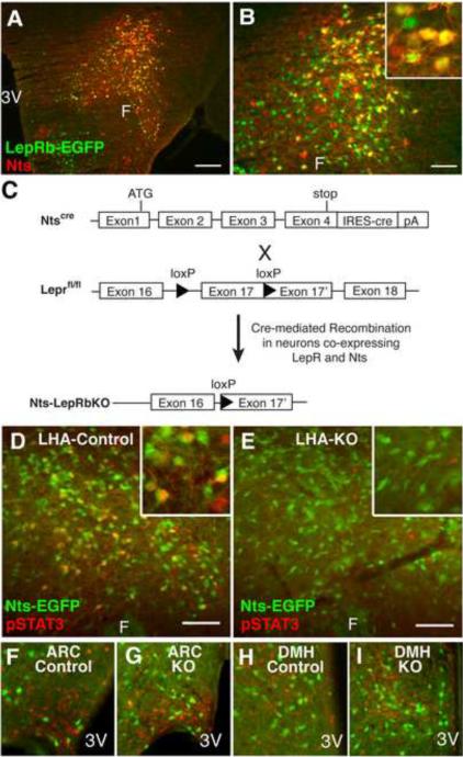
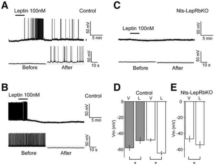
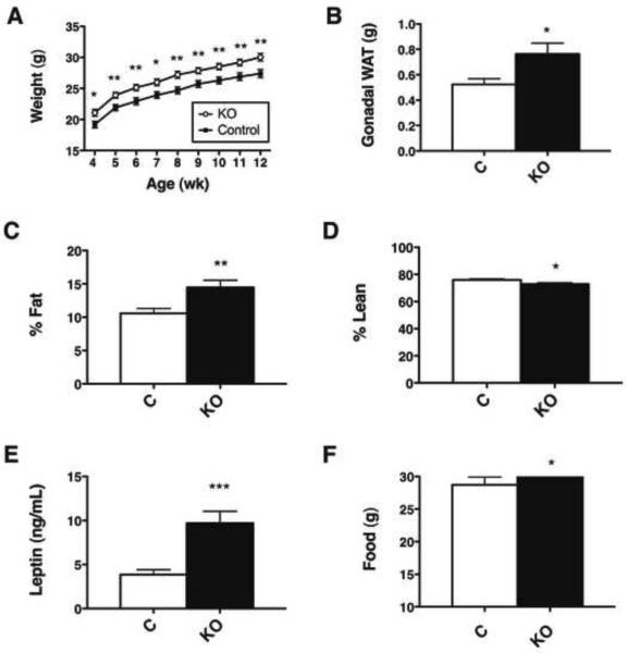
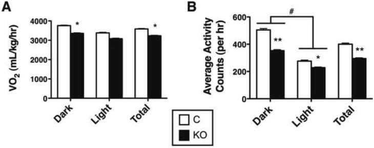
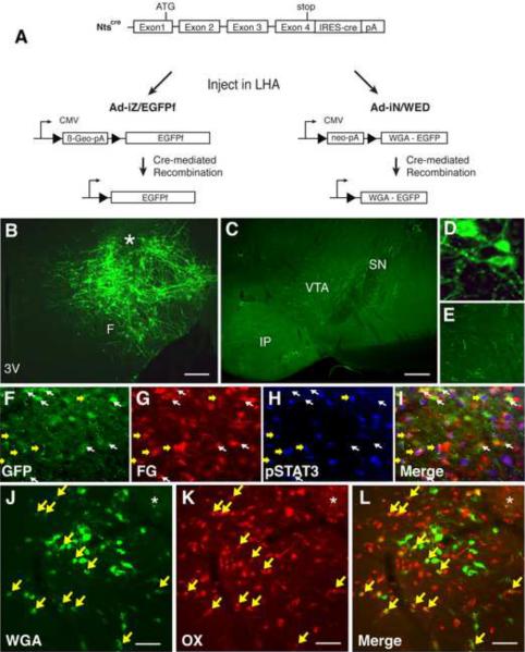
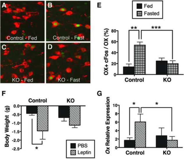
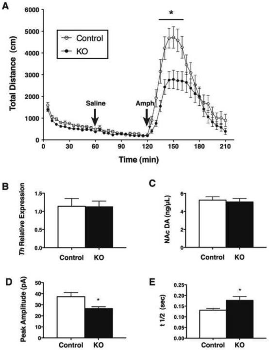
Leptin acts on leptin receptor (LepRb)-expressing neurons throughout the brain, but the roles for many populations of LepRb neurons in modulating energy balance and behavior remain unclear. We found that the majority of LepRb neurons in the lateral hypothalamic area (LHA) contain neurotensin (Nts). To investigate the physiologic role for leptin action via these LepRb(Nts) neurons, we generated mice null for LepRb specifically in Nts neurons (Nts-LepRbKO mice). Nts-LepRbKO mice demonstrate early-onset obesity, modestly increased feeding, and decreased locomotor activity. Furthermore, consistent with the connection of LepRb(Nts) neurons with local orexin (OX) neurons and the ventral tegmental area (VTA), Nts-LepRbKO mice exhibit altered regulation of OX neurons and the mesolimbic DA system. Thus, LHA LepRb(Nts) neurons mediate physiologic leptin action on OX neurons and the mesolimbic DA system, and contribute importantly to the control of energy balance.


Figures

Figure 1
Figure 1. A Subpopulation of LHA LepRb neurons contains Nts
(A) Immunofluorescent detection of GFP (green) and Nts (red) in colchicine-treated LepRbEGFP animals. (B) Enlargement of (A). (C) Schematic diagram depicting the generation of Nts-LepRbKO mice lacking functional LepRb only in Nts neurons. (D–I) Ntscre mice were bred onto the Rosa26-EGFP reporter line so that cre induces the expression of EGFP to identify cre-containing (Nts) neurons. Nts-EGFP (Control) and Nts-LepRbKO-EGFP (KO) mice (n=4 each) were treated with leptin (IP, 5 mg/kg, 2h) and analyzed via immunohistochemistry for pSTAT3 (red nuclei, pseudocolored) and immunofluoresence for EGFP. (D) LHA of a leptin-treated control mouse. (E) LHA of a leptin-treated KO mouse. (F, G) Leptin-induced pSTAT3 is similar in the ARC of control (F) and KO mice (G). (H, I) Leptin-induced pSTAT3 is similar in the DMH of control (H) and KO mice (I). Insets: higher magnification view of labeled LHA neurons. Scale bar = 100 μm. 3V= third cerebral ventricle; F = fornix.
Figure 2
Figure 2. Electrophysiology of LHA Nts Neurons in Response to Leptin
Brain slice recordings of EGFP (i.e., Nts) neurons in the LHA of Nts-EGFP (Control) and Nts-LepRbKO-EGFP (Nts-LepRbKO) mice treated with vehicle (V) or leptin (100 nM, L). Representative traces (A, B, C; bottom panel, on the expanded time scale) and aggregate responses (D, E) from leptin-depolarized (A, D-grey bars) and leptin-hyperpolarized neurons (B, D- white bars) in Control mice, and (C, E) all recorded neurons in Nts-LepRbKO mice. All the recordings were performed in the presence of GABAA, glycine and glutamate receptor antagonists. Data represent mean ± SEM, * = p<0.01 by paired t test.
Figure 3
Figure 3. Leptin action via LHA LepRbNts neurons regulates body weight and adiposity via
(A) Body weight, (B) gonadal fat pad weight, (C) percentage fat mass, and (D) percentage lean mass of male Control and Nts-LepRbKO (KO) mice (12–14 wk old, n=23–25 per genotype). (E) ELISA analysis of terminal serum for leptin (13–15 wk old, KO n=15, control n = 16). (F) Weekly food intake from 4–5 wk of age. (n=25 per genotype). Graphed data represent average values ± SEM. *p<0.05 by Student's t test.
Figure 4
Figure 4. Leptin action via LHA LepRbNts neurons regulates VO2 and Locomotor Activity
12–14 week old male Nts-LepRbKO (KO) and Control mice (KO n=20, control n = 16) were subjected to CLAMS analysis to determine (A) VO2 and (B) ambulatory locomotor activity. Data are shown for dark cycle (Dark), light cycle (Light) and averaged over 24 hours (Total). Graphed data represent average values ± SEM. ANOVA: *p < 0.05, **p < 0.01 and ***p < 0.001 for Control vs. KO; # p<0.05 for light vs. dark.
Figure 5
Figure 5. LHA Nts neurons project to the VTA and within the LHA onto OX neurons
(A) Schematic diagram showing cre-mediated expression of EGFPf and WGA in infected Nts-expressing neurons following stereotaxic injection of Ad-iZ/EGFPf or Ad-IN/WED, respectively, into the LHA of Ntscre mice. (B) Immunofluorescent detection of EGFPf in the LHA of Ntscre mice following intra-LHA injection of Ad-iZ/EGFPf identifies cell bodies and fibers (projections) in the LHA. (C) Immunofluorescent detection of EGFPf in the VTA of Ntscre mice after intra-LHA injection of Ad-iZ/EGFPf. (D, E) Enlargements of B and D, respectively. (F–I) LHA images from leptin-treated (2h, IP, 5 mg/kg) NtsEGFP mice injected with the retrograde tracer FG in the VTA. (F) GFP-IR, (G) FG-IR, (H) pSTAT3-IR, and (I) merged image. Yellow arrows indicate neurons positive for pSTAT3, FG and GFP. White arrows identify GFP+FG neurons that do not contain pSTAT3. (J–L) LHA from an Ntscre mouse injected with Ad-IN/WED. (J) Immunofluoresence for WGA (green), (K) OX (red), and (L) merged. Yellow arrows: neurons that contain OX + WGA. Scale bars in (B) = 100 μm. F= fornix; 3V = third cerebral ventricle; VTA = ventral tegmental area; SN = substantia nigra (pars compacta); IP= interpeduncular tubercle; * = injection site.
Figure 6
Figure 6. LHA LepRbNts neurons regulate OX neurons
Adult (12–15 wk) male Nts-LepRbKO (KO) and Control mice were ad libitum fed (A, C) or fasted for 26h (B, D) and analyzed by immunohistochemistry for cFos (green nuclei, pseudocolored) and immunofluoresence of OX neurons (red). (E) Percentage of OX neurons containing cFos in fed and fasted Control and KO mice. Control fed n= 4, control fasted n=5, KO fed n=6, KO fasted n=7. (F, G) Adult male Control and KO mice were treated with PBS or leptin (IP, 5 mg/kg) every 12h for 26 h total for the assessment of body weight and Ox expression. Control + PBS n=12, control + leptin n=10, KO + PBS n=12, KO + PBS n=15. (F) Weight change over 26h of treatment in control and KO mice. (G) Ox expression in the LHA of control and KO mice. Data are plotted as mean ± SEM; * p<0.05 via one-way ANOVA with Bonferroni post test.
Figure 7
Figure 7. LHA LepRbNts neurons modulate the mesolimbic DA system
(A) Adult male Nts-LepRbKO (KO) and littermate Control mice were analyzed for AMPH-induced locomotor activity. Mice were placed in the activity monitoring chamber at time 0, given an IP saline injection at 60 min, AMPH (4 mg/kg, IP) at 120 minutes, and were followed for an additional 90 minutes. Graphed data represent the average total distance (cm) ambulated ± SEM, control n=15, KO n=20. (B) Relative Th expression in the VTA and (C) DA in the NAc of control and KO mice. Data are plotted as the average ± SEM, control n=12, LeprNtsKO n=15. (D, E) Mean evoked dopamine spike amplitude (D) and t1/2 (E) in the NAc shell in KO and Control mice. Amplitude: KO, n=36 stimulations from 8 slices in 5 animals; Control, n=42 stimulations from 9 slices in 5 animals. t1/2: KO, n=36 stimulations from 8 slices in 5 animals; Control n=42 stimulations from 9 slices in 5 animals. Data are plotted as the average ± SEM, *p < 0.05 by ANOVA.

References

    1. Anaclet C, Parmentier R, Ouk K, Guidon G, Buda C, Sastre JP, Akaoka H, Sergeeva OA, Yanagisawa M, Ohtsu H, et al. Orexin/hypocretin and histamine: distinct roles in the control of wakefulness demonstrated using knock-out mouse models. J Neurosci. 2009;29:14423–14438. - PMC - PubMed
    1. Aston-Jones G, Smith RJ, Moorman DE, Richardson KA. Role of lateral hypothalamic orexin neurons in reward processing and addiction. Neuropharmacology. 2009;56(Suppl 1):112–121. - PMC - PubMed
    1. Balthasar N, Coppari R, McMinn J, Liu SM, Lee CE, Tang V, Kenny CD, McGovern RA, Chua SC, Jr., Elmquist JK, et al. Leptin Receptor Signaling in POMC Neurons Is Required for Normal Body Weight Homeostasis. Neuron. 2004;42:983–991. - PubMed
    1. Banks AS, Davis SM, Bates SH, Myers MG., Jr. Activation of downstream signals by the long form of the leptin receptor. J Biol Chem. 2000;275:14563–14572. - PubMed
    1. Berthoud HR. Interactions between the “cognitive” and “metabolic” brain in the control of food intake. Physiology & behavior. 2007;91:486–498. - PubMed

Publication types

MeSH terms