. 2011 Sep 7;14(3):428-34.
doi: 10.1016/j.cmet.2011.07.010.

Mutations in MTFMT underlie a human disorder of formylation causing impaired mitochondrial translation

Affiliations

Mutations in MTFMT underlie a human disorder of formylation causing impaired mitochondrial translation

Elena J Tucker et al. Cell Metab. .

Abstract

The metazoan mitochondrial translation machinery is unusual in having a single tRNA(Met) that fulfills the dual role of the initiator and elongator tRNA(Met). A portion of the Met-tRNA(Met) pool is formylated by mitochondrial methionyl-tRNA formyltransferase (MTFMT) to generate N-formylmethionine-tRNA(Met) (fMet-tRNA(met)), which is used for translation initiation; however, the requirement of formylation for initiation in human mitochondria is still under debate. Using targeted sequencing of the mtDNA and nuclear exons encoding the mitochondrial proteome (MitoExome), we identified compound heterozygous mutations in MTFMT in two unrelated children presenting with Leigh syndrome and combined OXPHOS deficiency. Patient fibroblasts exhibit severe defects in mitochondrial translation that can be rescued by exogenous expression of MTFMT. Furthermore, patient fibroblasts have dramatically reduced fMet-tRNA(Met) levels and an abnormal formylation profile of mitochondrially translated COX1. Our findings demonstrate that MTFMT is critical for efficient human mitochondrial translation and reveal a human disorder of Met-tRNA(Met) formylation.


Figures

Figure 1
Figure 1. Combined OXPHOS deficiency due to a defect in mitochondrial translation
(A) Biochemical analysis of OXPHOS complexes relative to citrate synthase (CS) in fibroblasts (Fb), muscle (M) or liver (L), expressed as a % of mean from healthy controls. (B) SDS-PAGE analysis of 35S-methionine-labeled mtDNA-encoded proteins from control and patient fibroblasts. MtDNA-encoded subunits of complex I (ND1, ND2, ND3, ND5, ND4L), complex III (cytb), complex IV (COX1, COX2, COX3) and complex V (ATP6, ATP8) are shown. (C) Gel in (B) was immunoblotted with antibodies against mtDNA-encoded ND1, COX1, COX2 and nuclear-encoded SDHA (complex II; loading control). See also Supplementary Figure S1.
Figure 2
Figure 2. Identification of pathogenic compound heterozygous mutations in MTFMT
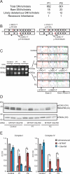
(A) Number of MitoExome variants that pass prioritization filters. (B) Schematic diagram of MTFMT showing the location of mutations in P1 and P2 (red bars), exon skipping (gray boxes), and primers for RT-PCR (forward and reverse arrows). (C) Electrophoresis of RT-PCR products demonstrates a smaller cDNA species (280bp) in P1 and P2 that is particularly prominent in cells grown in the presence of cycloheximide (+CHX). Top: Sequence chromatograms of full-length MTFMT RT-PCR products (-CHX) to confirm compound heterozygosity. Bottom: Sequence chromatograms of the smaller RT-PCR products (+CHX) shows patient cDNA lacks the c.382C>T (P1) or c.374C>T (P2) mutations and skips exon 4, which carries the shared c.626C>T mutation. (D-E) Patient and control fibroblasts were transduced with MTFMT cDNA or control C8orf38 cDNA (D) Representative SDS-PAGE western blot shows reduced COX2 and NDUFB8 in patient fibroblasts and restoration of protein levels with MTFMT but not C8orf38 transduction. The 70kDa complex II subunit acts as a loading control. (E) Protein expression was quantified by densitometry and bar charts show the level of complex I (NDUFB8) or complex IV (COX2) relative to complex II (70kDa) normalized to control, before and after transduction. Bars show the mean of 3 biological replicates and error bars indicate ± 1 s.e.m. Asterisks indicate p<0.05 (*), p<0.01 (**) and p<0.001(***). See also Supplementary Figure S2.
Figure 3
Figure 3. Patient fibroblasts have a defect in Met-tRNAMet formylation
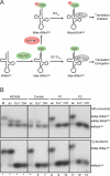
(A) In metazoan mitochondria, a single tRNAMet species acts as both initiator and elongator tRNAMet. After aminoacylation of tRNAMet by the mitochondrial methionyl-tRNA synthetase (MetRSmt), a portion of Met-tRNAMet is formylated by MTFMT to generate fMet-tRNAMet. fMet-tRNAMet is used by the mitochondrial IF2 (IF2mt) to initiate translation, whereas Met-tRNAMet is recognized by the mitochondrial EF-Tu (EF-Tumt) for the elongation of translation products. (B) Total RNA from control (lanes 5 – 7) and patient fibroblasts (P1: lanes 8 – 10; P2: lanes 11 – 13) was separated by acid-urea PAGE. Total RNA from MCH58 cells is shown as a reference (lanes 1 – 4). The mitochondrial tRNAMet (top panel) and the cytoplasmic initiator tRNAiMet (bottom panel) were detected by Northern hybridization. Total RNA was isolated under acidic conditions, which preserves both the Met-tRNAMet and fMet-tRNAMet (ac); tRNAs were treated with copper sulfate (Cu2+), which specifically deacylates Met-tRNAMet, but not fMet-tRNAMet; or with base (OH-) which deacylates both Met-tRNAMet and fMet-tRNAMet. Base treated tRNA was re-aminoacylated in vitro with Met using MetRS generating a Met-tRNAMet standard (M).
Figure 4
Figure 4. Analysis of the COX1 N-terminus in patient fibroblasts
(A-C) Annotated MS/MS spectra confirming correct targeting of the three possible N-termini of COX1. The sequence of the peptide is MFADRWLFSTNHK where the Met residue may be (A) formylated, (B) unformylated , or (C) absent (des-Met). The N-terminal amino acid of the peptide is shown in bold. The sequence ladder spacing corresponds to the b-ion series, and y-ion series fragmentation positions are shown below. fM=formyl-Methionine. Insets show high-resolution, high mass accuracy precursors from which the fragmentation spectra were derived. Given their sequence similarity, peptides are expected to have similar ionization efficiencies. (D) Extracted ion chromatograms (XICs) of three N-terminal states of COX1 ([fMet,Met,des-Met]FADRWLFSTNHK), normalized to an internal COX1 peptide (VFSWLATLHGSNMK). (E) Fractional ion current of the three N-terminal states of COX1 from immunoprecipitated complex IV of patients and controls. See also Supplementary Figure S3.

References

    1. Anderson S, Bankier AT, Barrell BG, de Bruijn MH, Coulson AR, Drouin J, Eperon IC, Nierlich DP, Roe BA, Sanger F, et al. Sequence and organization of the human mitochondrial genome. Nature. 1981;290:457–465. - PubMed
    1. Bentley DR, Balasubramanian S, Swerdlow HP, Smith GP, Milton J, Brown CG, Hall KP, Evers DJ, Barnes CL, Bignell HR, et al. Accurate whole human genome sequencing using reversible terminator chemistry. Nature. 2008;456:53–59. - PMC - PubMed
    1. Calvo SE, Tucker EJ, Compton AG, Kirby DM, Crawford G, Burtt NP, Rivas M, Guiducci C, Bruno DL, Goldberger OA, et al. High-throughput, pooled sequencing identifies mutations in NUBPL and FOXRED1 in human complex I deficiency. Nature Genetics. 2010;42:851–858. - PMC - PubMed
    1. Durbin RM, Abecasis GR, Altshuler DL, Auton A, Brooks LD, Gibbs RA, Hurles ME, McVean GA. A map of human genome variation from population-scale sequencing. Nature. 2010;467:1061–1073. - PMC - PubMed
    1. Enriquez JA, Attardi G. Analysis of aminoacylation of human mitochondrial tRNAs. Methods Enzymol. 1996;264:183–196. - PubMed

Publication types

MeSH terms