. 2011 Nov;152(11):2564-2574.
doi: 10.1016/j.pain.2011.07.020. Epub 2011 Sep 9.

Preventive or late administration of anti-NGF therapy attenuates tumor-induced nerve sprouting, neuroma formation, and cancer pain

Affiliations

Preventive or late administration of anti-NGF therapy attenuates tumor-induced nerve sprouting, neuroma formation, and cancer pain

Juan Miguel Jimenez-Andrade et al. Pain. 2011 Nov.

Abstract

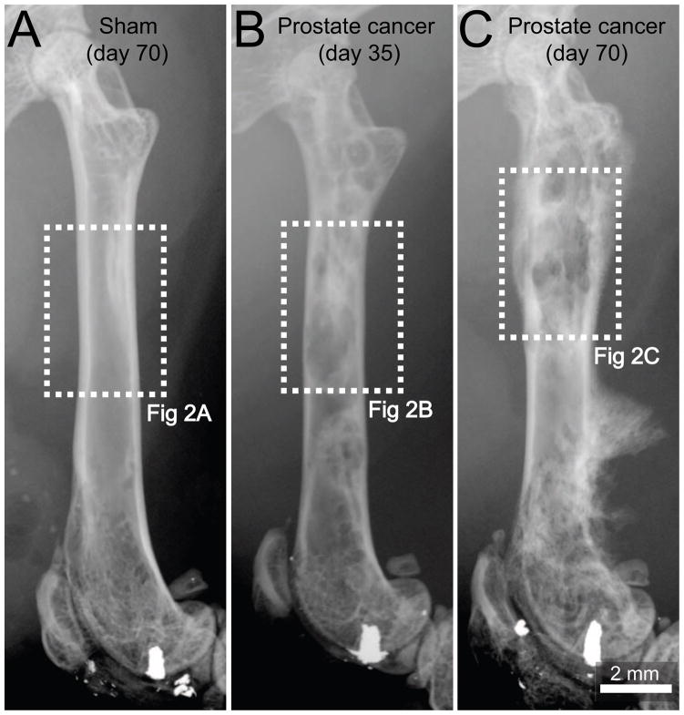
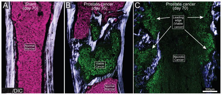
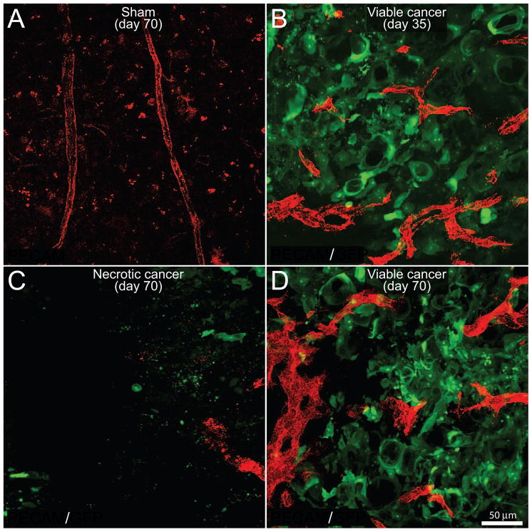
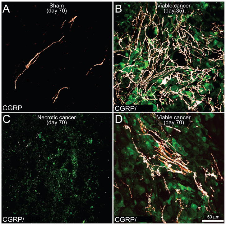
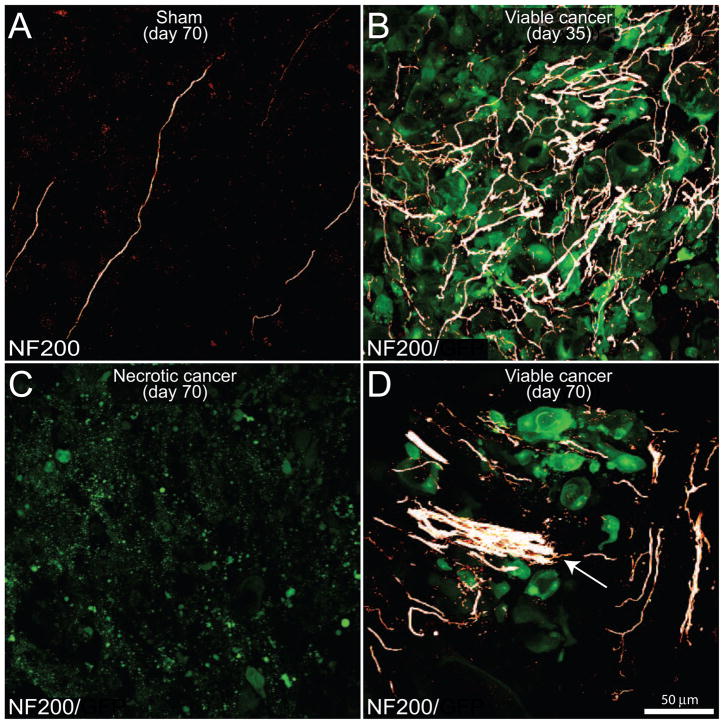
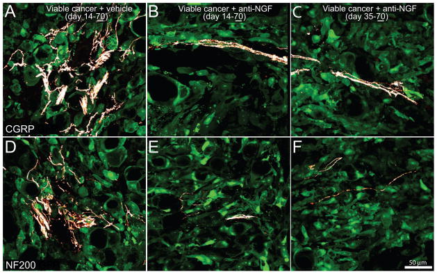
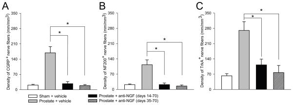
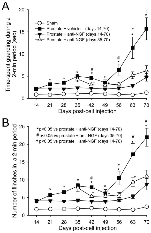
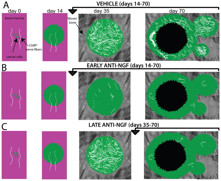
Early, preemptive blockade of nerve growth factor (NGF)/tropomyosin receptor kinase A (TrkA) attenuates tumor-induced nerve sprouting and bone cancer pain. A critical unanswered question is whether late blockade of NGF/TrkA can attenuate cancer pain once NGF-induced nerve sprouting and neuroma formation has occurred. By means of a mouse model of prostate cancer-induced bone pain, anti-NGF was either administered preemptively at day 14 after tumor injection when nerve sprouting had yet to occur, or late at day 35, when extensive nerve sprouting had occurred. Animals were humanely killed at day 70 when, in vehicle-treated animals, significant nerve sprouting and neuroma formation was present in the tumor-bearing bone. Although preemptive and sustained administration (days 14-70) of anti-NGF more rapidly attenuated bone cancer nociceptive behaviors than late and sustained administration (days 35-70), by day 70 after tumor injection, both preemptive and late administration of anti-NGF significantly reduced nociceptive behaviors, sensory and sympathetic nerve sprouting, and neuroma formation. In this model, as in most cancers, the individual cancer cell colonies have a limited half-life because they are constantly proliferating, metastasizing, and undergoing necrosis as the parent cancer cell colony outgrows its blood supply. Similarly, the sensory and sympathetic nerve fibers that innervate the tumor undergo sprouting at the viable/leading edge of the parent tumor, degenerate as the parent cancer cell colony becomes necrotic, and resprout in the viable, newly formed daughter cell colonies. These results suggest that preemptive or late-stage blockade of NGF/TrkA can attenuate nerve sprouting and cancer pain.


Conflict of interest statement

Conflicts of interest statement

None of the authors of this study claim a conflict of interest.

Figures

Figure 1
Figure 1. Pathological bone remodeling induced by prostate cancer cells growing within the femur
Representative radiographic images of a mouse femur at day 70 following injection of culture medium alone (sham, A), at day 35 following injection of ACE-1 prostate cancer cells (B) or day 70 following injection of prostate cancer cells (C). At day 35 post-cancer cell injection the tumor-injected mouse femur shows osteoblastic lesions, characterized by pathological bone formation in the intramedullary space, which generate diaphyseal bridging structures (B). Note that by day 70, the osteoblastic lesions increase in magnitude and an aggressive periosteal reaction is evidenced as a dense and disorganized appearance of the cortical bone in the distal methaphysis and formation of a Codman’s triangle-like structure in the distal diaphysis (C). A Codman’s triangle develops when a portion of periosteum is lifted off of the cortex by tumor and has been reported to occur in bones from patients with metastatic prostate cancer. Sham-injected femurs show no evidence of bone formation or bone destruction (A). Dashed line rectangles in white represent the areas of the bones illustrated in figure 2.
Figure 2
Figure 2. The evolving histopathology as prostate cancer cells grow within the mouse femur
Differential interphase contrast (DIC) images overlaid on confocal green fluorescent protein (GFP) images (20 μm-thick) of a sham (A) and prostate cancer cell-injected femur from mice sacrificed at days 35 (B) and 70 (C) post-cancer cell injection. DIC images were acquired to visualize cortical and pathological woven bone. As the GFP+ tumor cells (green) grow in normal bone marrow (normal tightly packed hematopoietic cells, pink) they induce the formation of woven bone around the tumor cell colony and eventually form an osteoblastic lesion (B). As tumor disease progresses (day 70), the parent cancer cell colonies undergo necrosis, whereas viable, daughter cancer cell colonies, which are also surrounded by new bone, form at sites more distant from bone marrow (C). Note that at late stages of the disease, the diameter of the femur is greater than that of the femur at day 35 post-tumor injection, or sham bones due to the continuous bone growth induced by prostate cancer cells. Note that osteoblastic lesions are not present in the sham femur (A). For illustration purposes, the background signal which corresponds to healthy bone marrow is presented as the color pink and the GFP+ tumor cells as green.
Figure 3
Figure 3. Newly formed prostate cancer cell colonies are highly viable and well vascularized whereas older cancer cell colonies lose their vascular supply and undergo necrosis and death
Confocal images of bone sections (20 μm-thick) from sham mice (A) or mice sacrificed at days 35 (B) or 70 (C,D) post-tumor cell injection. GFP+ cancer cell (green)-bearing bone sections were immunostained with an antibody against the platelet endothelial cell adhesion molecule (PECAM, red) to immunolabel endothelial cells of blood vessels. Blood vessels in the bone marrow in naïve (data not shown) and sham injected animals (A) have a highly organized, homogenous and mostly linear morphology. As GFP+ prostate tumor cells grow within the bone (B), there is an increase in the density of PECAM+ blood vessels (red) inside and immediately surrounding the cancer cell colonies. Note that these PECAM+ blood vessels have a highly heterogeneous and disorganized morphology as compared to blood vessels in the bone marrow of sham mice (B). At day 70, the older cancer cell colonies show a loss of PECAM+ blood vessels and as well as GFP+ cancer cells (C), whereas the immediately adjacent daughter cancer colonies are characterized by a high density of PECAM+ blood vessels and GFP+ cancer cells (D). Images were acquired at the metaphyseal region of the bone marrow and were projected from 40 optical sections at 0.5 μm intervals.
Figure 4
Figure 4. The evolving reorganization of CGRP+ sensory nerve fibers with disease progression
Confocal images of bone sections (20 μm-thick) from sham mice (A) or mice sacrificed at days 35 (B) or 70 (C,D) post prostate tumor cells injection. GFP+ cancer cell (green)-bearing bone sections were immunostained with an antibody against calcitonin gene-related peptide (CGRP, a marker of peptide-rich sensory nerve fibers, white). Note that in the sham mice (A), CGRP+ nerve fibers that innervate the healthy marrow space appear as single nerve fibers with a highly linear morphology. In contrast, as GFP+ prostate tumor cells proliferate and form tumor colonies (day 35 post-cell injection; B), the CGRP+ sensory nerve fibers undergo marked sprouting as characterized by an increased density, highly branched architecture, and disorganized morphology as compared to nerve fibers innervating the marrow space in sham animals (A). At day 70 post-cell injection the older parent cancer cell colonies show signs of necrosis as evidenced by a loss of GFP expression in the cancer cells, as well as a decrease in the density of CGRP+ nerve fibers (C), whereas adjacent newly formed daughter cancer colonies show robust sprouting and formation of neuroma-like structures by CGRP+ nerve fibers (D). Images were acquired at the metaphyseal region of the bone marrow and were projected from 40 optical sections at 0.5 μm intervals.
Figure 5
Figure 5. The evolving reorganization of NF200+ sensory nerve fibers as prostate cancer cells proliferate, metastasize and undergo necrosis in the mouse femur
Confocal images of bone sections (20 μm-thick) from sham mice (A) or mice sacrificed at days 35 (B) or 70 (C,D) post-injection. GFP+ cancer cell (green)-bearing bone sections were incubated with an antibody against 200 kD neurofilament (NF200, a marker of myelinated nerve fibers; white). Note that in the sham mice (A), NF200+ nerve fibers that innervate the healthy marrow space appear as single nerve fibers with a highly linear morphology. In contrast, as GFP+ prostate tumor cells proliferate and form tumor colonies at day 35 post-cell injection (B), the NF200+ sensory nerve fibers undergo marked sprouting characterized by a highly branched morphology and increased density as compared to sham mice. With time, older parent colonies show a dramatic loss of GFP+ cancer cells, as well as a decrease in the density of NF200+ nerve fibers (C), whereas immediately adjacent new daughter cancer cell colonies show robust expression of GFP as well as sprouting and formation of neuroma-like structures (arrow) by NF200 nerve fibers (D). Images were acquired at the metaphyseal region of the bone marrow and were projected from 40 optical sections at 0.5 μm intervals.
Figure 6
Figure 6. Sustained treatment with anti-NGF, given either preemptively or late in disease progression, decreases tumor-induced sprouting of CGRP+ and NF200+ sensory nerve fibers
Representative confocal images of viable cancer cell colonies of bones harvested from mice sacrificed at day 70 post-prostate tumor cell injection (underline of 70). With advanced disese progression prostate cancer cells (which are transfected with green fluorescent protein: GFP, green) form new viable colonies of cancer cells, in which there is dramatic sprouting and formation of neuroma-like structures by CGRP+ (A) and NF200+ (D) nerve fibers. Preemptive, sustained sequestration of NGF (10 mg/kg; i.p., administered from day 14 to day 70) or late administration (given from day 35 to day 70 post-cancer cell injection) significantly reduces the pathological tumor-induced sprouting and formation of neuroma-like structures of sensory CGRP+ (B, C) and NF200+ (E, F) nerve fibers at day 70 post-tumor injection. Images were acquired at the metaphyseal region of the bone marrow and were projected from 40 optical sections at 0.5 μm intervals.
Figure 7
Figure 7. Histograms showing that sustained administration of NGF sequestering therapy, given either early or late in disease progression, reduces prostate-induced nerve sprouting of CGRP+, NF200+, and TrkA+ nerve fibers
At day 70 post cell injection, the density of CGRP+ (A), NF200+ (B), and TrkA+ (C) nerve fibers is significantly greater in prostate + vehicle-treated mice compared to sham + vehicle-treated mice. At day 70 post-tumor injection, tumor-induced nerve sprouting is significantly attenuated by preemptive/sustained administration or anti-NGF (10 mg/kg; i.p., given from day 14 to day 70 post-cell injection) or by late/sustained administration of anti-NGF (10 mg/kg; i.p., given from day 35 to day 70 post-cell injection). Nerve fiber density was determined by measuring the total length of nerve fibers in areas where viable cancer cells were present. Brackets indicate the groups being compared. *p<0.05. Bars represent the mean ± SEM for at minimum six mice.
Figure 8
Figure 8. Anti-NGF treatment, when given early or late in disease progression, attenuates prostate cancer-induced nociceptive behaviors
Nociceptive behaviors including spontaneous guarding (A) and flinching (B) recorded in sham mice (needle placement + injection of culture medium), prostate cancer-bearing mice treated with vehicle, prostate cancer-bearing mice treated preemptively with anti-NGF (from days 14 to 70 post-cell injection) and prostate cancer mice treated late with anti-NGF (from days 35 to 70 post-cell injection). Note that nociceptive behaviors in tumor-bearing mice are evident by day 14 post-cell injection and are significantly greater than sham mice at all time points shown. Preemptive sustained treatment with anti-NGF which was commenced at day 14 post-tumor cell injection significantly decreased nociceptive behaviors. Importantly, late sustained treatment with anti-NGF which was commenced at day 35 post-tumor injection (when robust sprouting in the parent cell colonies has already occurred) also decreased the nociceptive behaviors. Anti-NGF was given every 5 days (10 mg/kg, i.p.) and each point represents the mean ± SEM.
Figure 9
Figure 9. Schematic illustrating the effect that preemptive or late administration of anti-NGF has on nerve fiber pathology with prostate cancer disease progression in bone
A) Following initial injection and confinement of the prostate tumor cells into the marrow space of the femur, there are small cancer cell colonies composed mainly of proliferating cancer cells that are innervated only by the sensory nerve fibers that normally innervate the bone, which have a linear and regular morphology. As the prostate tumor cells proliferate within the bone marrow (day 14) the tumor cells activate, injure and then destroy the very distal processes of sensory fibers that innervate the bone marrow (dashed lines). By day 35 post-tumor cell injection the cancer cells colonies (which are now composed of both cancer cells and their associate stromal cells) increase in size and are surrounded by copious amounts of newly formed woven bone, which has been induced by the prostate cancer colony. At this time point there is a clear remodeling of TrkA+ sensory and sympathetic nerve fibers and this reorganization is characterized by an increased density of nerve fibers which are highly branched and have a disorganized morphology that is never observed in the normal marrow space. At late stages of the disease (day 70) the sensory and sympathetic nerve fibers that innervate the viable, well vascularized daughter cell colonies show significant sprouting and neuroma formation whereas there is little evidence of any sensory or sympathetic nerve fibers innervating the older, now necrotic parent cancer cell colonies. Early and sustained anti-NGF therapy (day 14–70 day post-tumor cell injection) prevents sprouting and neuroma formation throughout the time the therapy is administered. In contrast, sustained anti-NGF treatment (day 35–70) prevents the nerve sprouting and neuroma formation which normally occur in the later formed daughter cell colonies, which are the only viable cell colonies that are present in the tumor-bearing bone at day 70 post-tumor injection.

Comment in

References

    1. Adriaenssens E, Vanhecke E, Saule P, Mougel A, Page A, Romon R, Nurcombe V, Le Bourhis X, Hondermarck H. Nerve growth factor is a potential therapeutic target in breast cancer. Cancer Res. 2008;68(2):346–351. - PubMed
    1. Ayala GE, Wheeler TM, Shine HD, Schmelz M, Frolov A, Chakraborty S, Rowley D. In vitro dorsal root ganglia and human prostate cell line interaction: redefining perineural invasion in prostate cancer. Prostate. 2001;49(3):213–223. - PubMed
    1. Black JA, Nikolajsen L, Kroner K, Jensen TS, Waxman SG. Multiple sodium channel isoforms and mitogen-activated protein kinases are present in painful human neuromas. Ann Neurol. 2008;64(6):644–653. - PubMed
    1. Bloom AP, Jimenez-Andrade JM, Taylor RN, Castaneda-Corral G, Kaczmarska MJ, Freeman KT, Coughlin KA, Ghilardi JR, Kuskowski MA, Mantyh PW. Breast Cancer-Induced Bone Remodeling, Skeletal Pain and Sprouting of Sensory Nerve Fibers. J Pain. 2011 - PMC - PubMed
    1. Bloom RA, Libson E, Husband JE, Stoker DJ. The periosteal sunburst reaction to bone metastases. A literature review and report of 20 additional cases. Skeletal Radiol. 1987;16(8):629–634. - PubMed

Publication types

MeSH terms