

Alterations in diffusion properties of white matter in Williams syndrome
- PMID: 21907520
- PMCID: PMC3199335
- DOI: 10.1016/j.mri.2011.07.012
Alterations in diffusion properties of white matter in Williams syndrome
Abstract
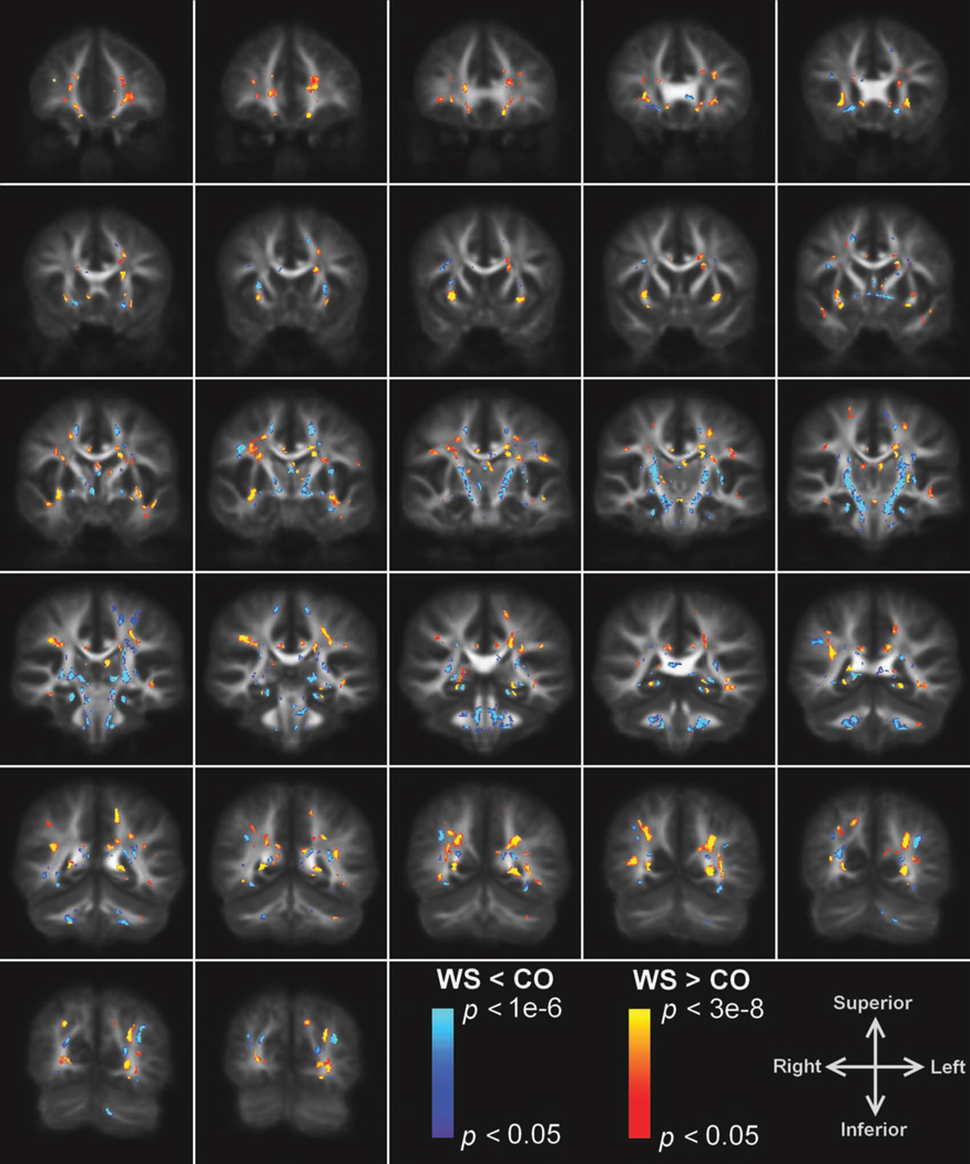
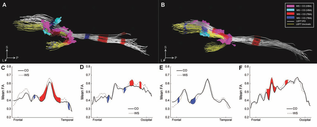
Diffusion tensor imaging (DTI) was used to investigate the involvement of brain white matter in Williams syndrome (WS), a genetic neurodevelopmental disorder. Whole-brain DTIs were obtained from 16 young adults with WS and 16 normal controls. A voxel-based analysis was performed to compare fractional anisotropy (FA) values between the two groups. A tract-based analysis was also performed to compare FA values between the two groups along two major white matter tracts that pass through the external capsule: the uncinate and inferior fronto-occipital fasciculi. Several regions of both increased and decreased FA were found within major white matter tracts that connect functional regions that have previously been implicated in the cognitive and neurological symptoms of the syndrome. The tract-based analysis provided additional insight into the involvement of specific white matter tracts implicated in the voxel-based analysis within the external capsule. The results from this study support previously reported changes in white matter diffusion properties in WS and demonstrate the potential usefulness for tract-based analysis in future studies of the disorder.
Copyright © 2011 Elsevier Inc. All rights reserved.
Figures
References
-
- Beuren AJ, Apitz J, Harmjanz D. Supravalvular aortic stenosis in association with mental retardation and a certain facial appearance. Circulation. 1962;26:1235–1240. - PubMed
-
- Williams JC, Barratt-Boyes BG, Lowe JB. Supravalvular aortic stenosis. Circulation. 1961;24:1311–1318. - PubMed
-
- Mervis CB, Robinson BF, Bertrand J, Morris CA, Klein-Tasman BP, Armstrong SC. The Williams syndrome cognitive profile. Brain Cogn. 2000;44(3):604–628. - PubMed
-
- Bellugi U, Adolphs R, Cassady C, Chiles M. Towards the neural basis for hypersociability in a genetic syndrome. Neuroreport. 1999;10(8):1653–1657. - PubMed
Publication types
MeSH terms
Grants and funding
LinkOut - more resources
Full Text Sources
