. 2011 Nov 1;174(2):202-10.
doi: 10.1016/j.ygcen.2011.08.019. Epub 2011 Sep 1.

Characterization of novel Pannexin 1 isoforms from rat pituitary cells and their association with ATP-gated P2X channels

Affiliations

Characterization of novel Pannexin 1 isoforms from rat pituitary cells and their association with ATP-gated P2X channels

Shuo Li et al. Gen Comp Endocrinol. .

Abstract

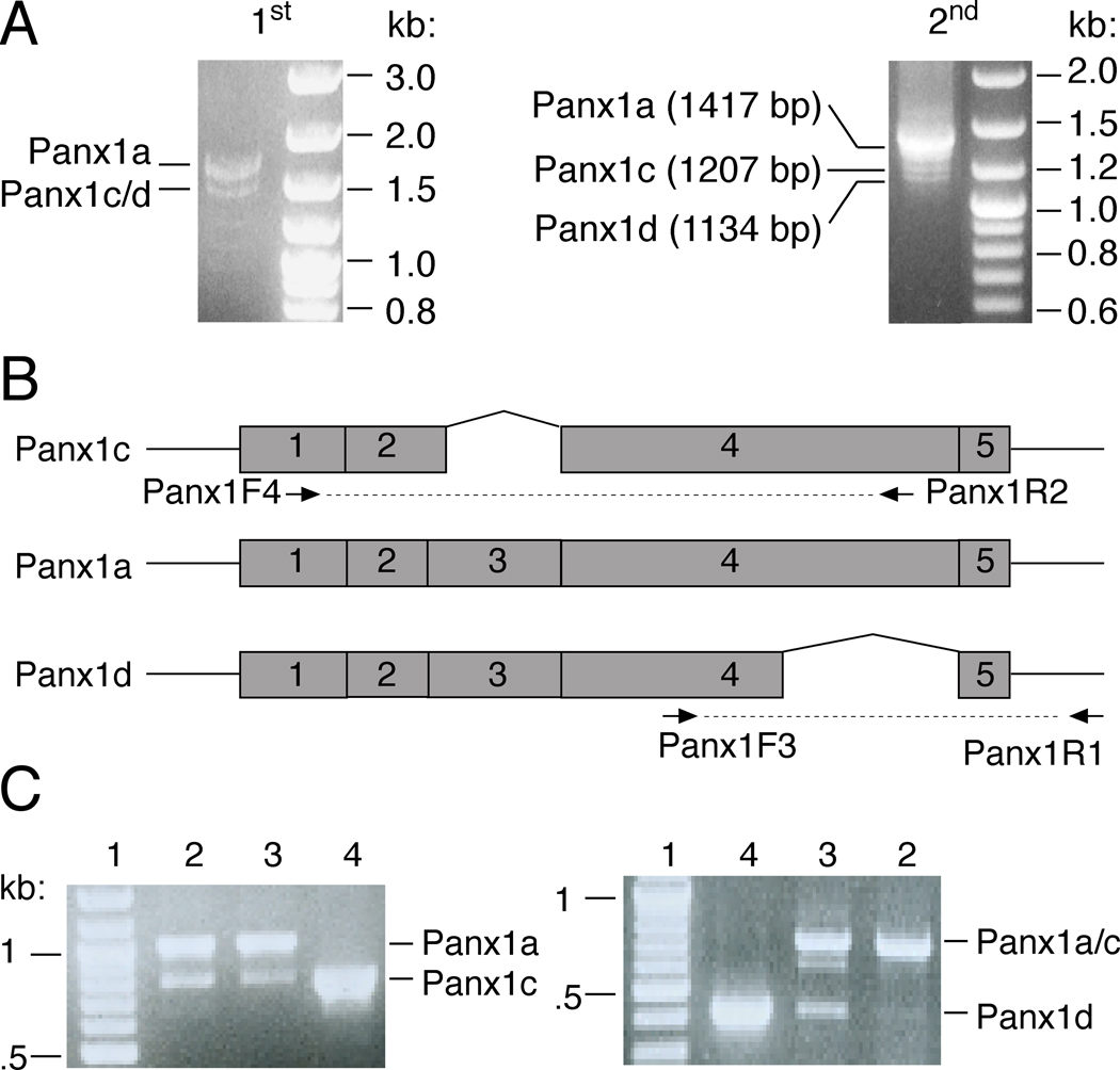
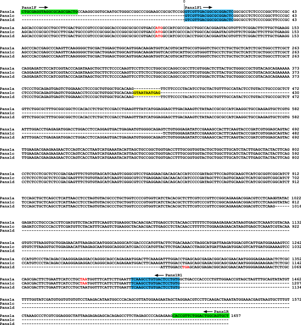
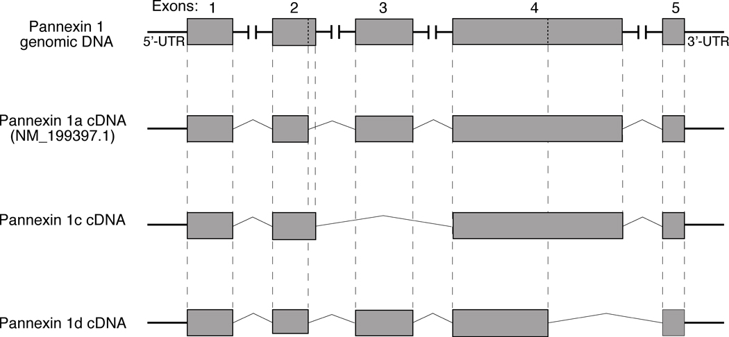
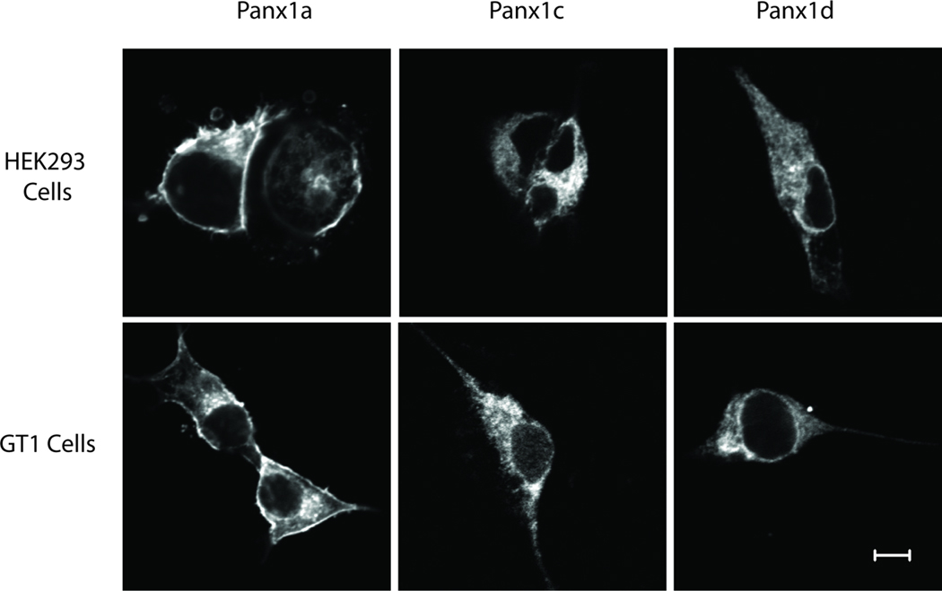
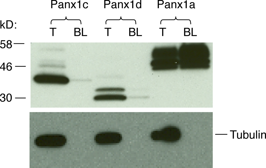
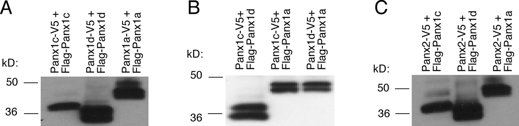
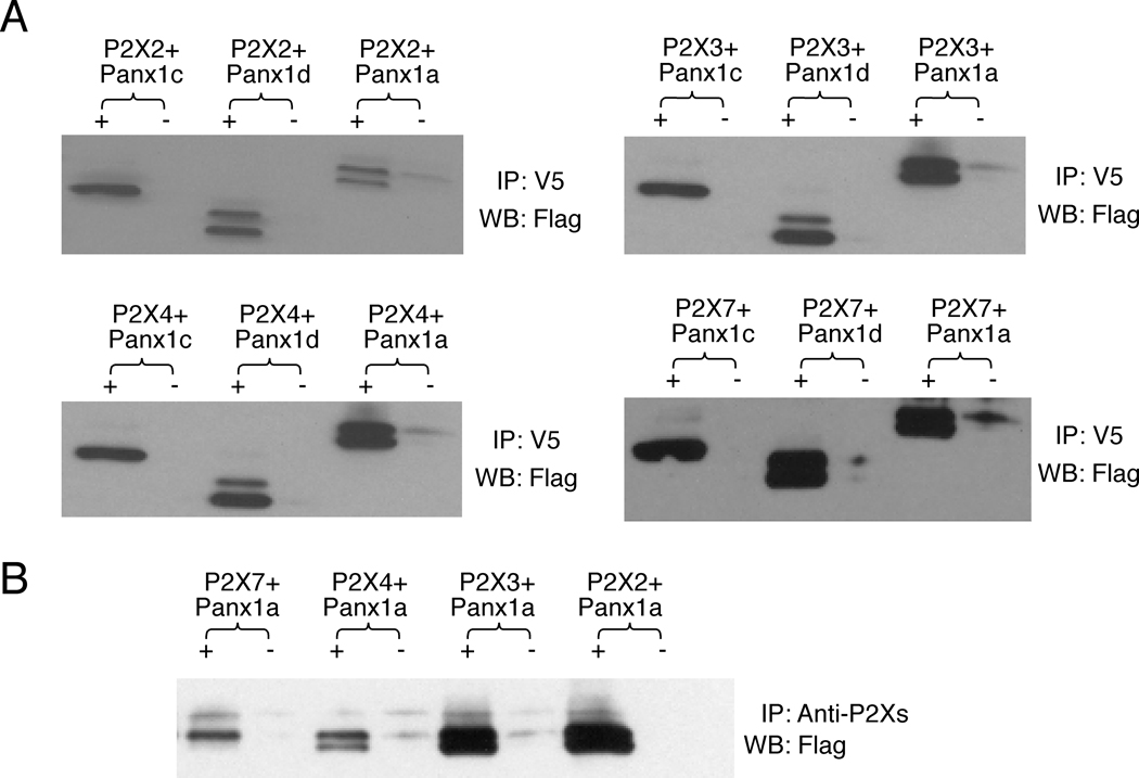
Our previous studies have showed that Pannexin 1 (Panx1), a member of a recently discovered family of gap junction proteins, is expressed in the pituitary gland. Here we investigated the presence and expression pattern of Panx1 isoforms in pituitary cells, their roles in ATP release, and their association with purinergic P2X receptor subtypes that are native to pituitary cells. In addition to the full-size Panx1, termed Panx1a, pituitary cells also express two novel shorter isoforms, termed Panx1c and Panx1d, which formation reflects the existence of alternative splicing sites in exons 2 and 4, respectively. Panx1c is lacking the Phe108-Gln180 sequence and P2X1d is missing the Val307-Cys426 C-terminal end sequence. Confocal microscopy and biotin labeling revealed that Panx1a is expressed in the plasma membrane, whereas Panx1c and Panx1d show the cytoplasmic localization when expressed as homomeric proteins. The three Panx1 isoforms and Panx2 form homomeric and heteromeric complexes in any combination. These splice forms can also physically associate with ATP-gated P2X2, P2X3, P2X4, and P2X7 receptor channels. The Panx1a-mediated ATP release in AtT-20 immortalized pituitary cells is attenuated when co-expressed with Panx1c or Panx1d. These results suggest that Panx1c and Panx1d may serve as dominant-negative effectors to modulate the functions of Panx1a through formation of heteromeric channels. The complex patterns of Panx1 expression and association could also define the P2X-dependent roles of these channels in cell types co-expressing both proteins.


Figures

Fig. 1
Fig. 1
Cloning and expression of the cDNAs for Panx1 isoforms from rat pituitary primary cells. (A) Nest RT-PCR was applied for amplification of the rat Panx1 cDNAs (A). 1st, the first round PCR products using primer pair Panx1F/Panx1R; 2nd, the second round nest PCR products using 100 times dilution of the first round PCR products as template and primer pair Panx1F1/Panx1R1. (B) Schematic representation of primer pairs used for detection of different Panx1 splicing variants. (C) The presence of Panx1c and Panx1d specific mRNA transcripts in normal and GH3 immortalized pituitary cells. The sizes of PCR products were as follows: 790 bp for Panx1c vs. 1.0 kb for Panx1a in detection of Panx1c isoform; 428 bp for Panx1d vs. 711 bp for Panx1a/c in detection of Panx1d isoform. 1: DNA marker; 2: GH3 cells cDNA; 3: pituitary cells cDNA; 4: plasmid control. Right panels show DNA markers.
Fig. 2
Fig. 2
Nucleotide sequences alignment of rat Panx1 isoforms. The primer pairs Panx1F/Panx1R (green) and Panx1F1/Panx1R1 (blue) were used in nest RT-PCR to amplify Panx1 cDNAs. The initiator ATG and stop codons are indicated in red. Yellow-labeled nucleotides indicate a Panx1c-specific sequence. The GenBank Accession numbers for Panx1c and Panx1d are GQ499839 and GQ499840, respectively.
Fig. 3
Fig. 3
Schematic representation of cDNA structures for Panx1 isoforms from rat pituitary cells. Top panel illustrates rat Panx1 genomic DNA. Putative translational exons in the genomic DNA are boxed in gray and horizontal lines illustrate four introns and 5’-UTR and 3’-UTR endings. Vertical dotted lines in exons 2 and 4 show the position of additional splicing sites for Panx1c and Panx1d, respectively. Bottom panels are schematic representation of cDNA for Panx1 splice variants. Vertical dashed lines show the corresponding regions between putative translational exons in the genomic DNA and Panx1 cDNAs.
Fig. 4
Fig. 4
Alignment of amino acid sequences of rat Panx1 isoforms. The deduced amino acid sequences of Panx1 isoforms are shown, starting with the initial methionine. The four predicted TM domains (I–IV) of Panx1 are marked in yellow. The arginine residue in position 75 that could contribute to ATP binding and inhibition of Panx1 currents is boxed. The three putative glycosylation sites (N204, N254 and N337) are indicated in blue. The conserved putative cysteine pairs (66/84 and 245/264) are indicated in green. The amino acid residues specific to Panx1c or Panx1d isoform are marked in red. Dashes indicate missing residues.
Fig. 5
Fig. 5
Cellular localization of Panx1 isoforms. HEK293 and GT1 cells were transiently transfected with FLAG-tagged Panx1a, Panx1c and Panx1d constructs. Cells were fixed, permeabilized, and fluorescence was analyzed 36 h after transfection using confocal laser scanning microscopy. Note that FLAG-tagged Panx1a, but not FLAG-tagged Panx1c and Panx1d isoforms, was expressed on the cell surface in both cell lines. Bar: 5 µm.
Fig. 6
Fig. 6
The biotin labeling analysis of the cell surface localization of Panx1 isoforms. HEK293 cells were transfected with N-terminal FLAG-tagged Panx1 constructs and 48 h later labeled with the membrane impermeable Sulfo-NHS-SS-Biotin. Total cell lysates (T) and biotin-labeled plasma membrane protein fractions (BL) were probed with anti-FLAG HRP-labeled antibody (top panel) and the same membranes were reprobed with anti-alpha tubulin antibody (controls, bottom panel). Note that only Panx1a was successfully biotinylated.
Fig. 7
Fig. 7
Formation of Panx homo- and hetero-complexes. HEK293 cells were co-transfected with FLAG- and V5-tagged (1:1) Panx1a, Panx1c and Panx1d constructs (A and B) or co-transfected with V5-tagged Panx2 and FLAG-tagged Panx1a, Panx1c or Panx1d constructs (C). Cell lysates were immunoprecipitated with anti-V5 antibody followed by immunoblotting using anti-FLAG HRP-labeled antibody. The protein size markers are shown on the center side.
Fig. 8
Fig. 8
Association of Panx1 proteins with P2XRs. HEK293 cells were co-transfected with FLAG- tagged Panx1a, Panx1c and Panx1d and V5-tagged P2X7R, P2X2R, P2X3R and P2X4R plasmids. Cell lysates were immunoprecipitated (IP) with anti-V5 antibody (A), anti-P2X2, P2X3, P2X4 or P2X7 antibodies (B) and immunoblotted with anti-FLAG HRP-labeled antibody (WB). +, IP with anti-V5 antibody (A) or anti-P2X2, P2X3, P2X4 and P2X7 antibodies (B); −, IP with normal mouse IgG (A) or normal rabbit serum (B) as negative controls.

References

    1. Bao L, Locovei S, Dahl G. Pannexin membrane channels are mechanosensitive conduits for ATP. FEBS Lett. 2004;572:65–68. - PubMed
    1. Baranova A, Ivanov D, Petrash N, Pestova A, Skoblov M, Kelmanson I, Shagin D, Nazarenko S, Geraymovych E, Litvin O, Tiunova A, Born TL, Usman N, Staroverov D, Lukyanov S, Panchin Y. The mammalian pannexin family is homologous to the invertebrate innexin gap junction proteins. Genomics. 2004;83:706–716. - PubMed
    1. Barbe MT, Monyer H, Bruzzone R. Cell-cell communication beyond connexins: the pannexin channels. Physiology (Bethesda) 2006;21:103–114. - PubMed
    1. Boassa D, Ambrosi C, Qiu F, Dahl G, Gaietta G, Sosinsky G. Pannexin1 channels contain a glycosylation site that targets the hexamer to the plasma membrane. J Biol Chem. 2007;282:31733–31743. - PubMed
    1. Bruzzone R, Barbe MT, Jakob NJ, Monyer H. Pharmacological properties of homomeric and heteromeric pannexin hemichannels expressed in Xenopus oocytes. J Neurochem. 2005;92:1033–1043. - PubMed

Publication types