Review
. 2011:62:109-41.
doi: 10.1016/B978-0-12-385952-5.00008-7.

Pharmacological chaperones for misfolded gonadotropin-releasing hormone receptors

Affiliations
Review

Pharmacological chaperones for misfolded gonadotropin-releasing hormone receptors

P Michael Conn et al. Adv Pharmacol. 2011.

Abstract

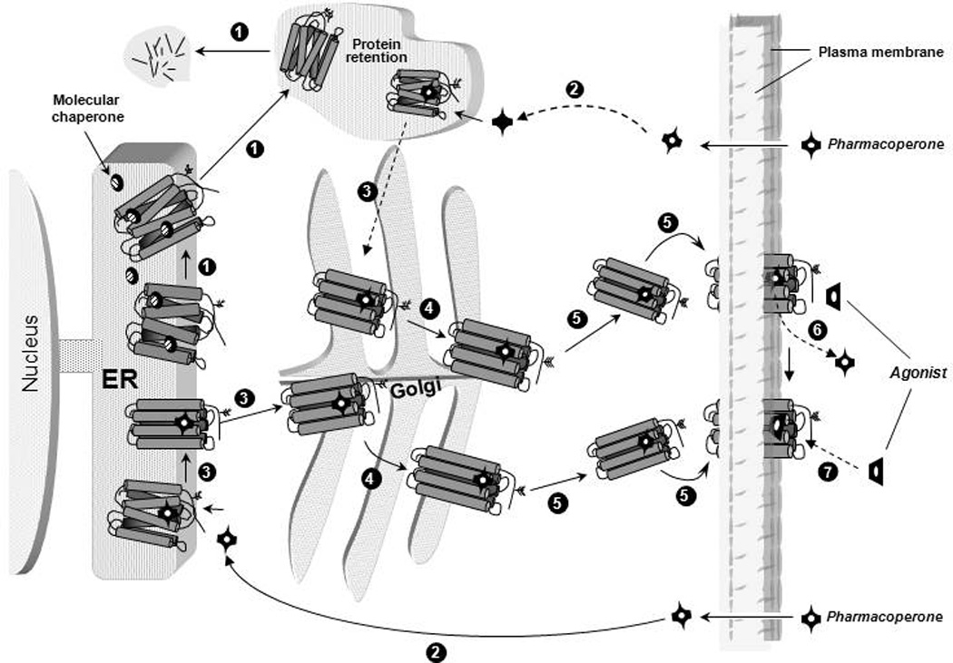
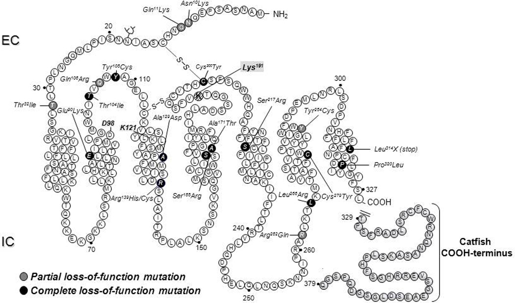
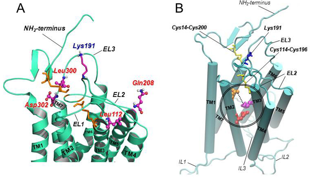
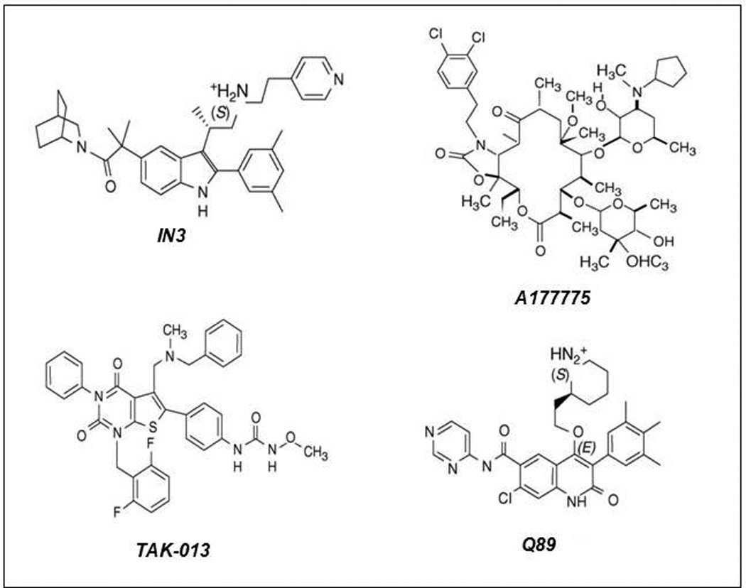
Structural alterations provoked by mutations or genetic variations in the gene sequence of G protein-coupled receptors (GPCRs) may lead to abnormal function of the receptor molecule. Frequently, this leads to disease. While some mutations lead to changes in domains involved in agonist binding, receptor activation, or coupling to effectors, others may cause misfolding and lead to retention/degradation of the protein molecule by the quality control system of the cell. Several strategies, including genetic, chemical, and pharmacological approaches, have been shown to rescue function of trafficking-defective misfolded GPCRs. Among these, pharmacological strategies offer the most promising therapeutic tool to promote proper trafficking of misfolded proteins to the plasma membrane (PM). Pharmacological chaperones or "pharmacoperones" are small compounds that permeate the PM, enter cells, and bind selectively to misfolded proteins and correct folding allowing routing of the target protein to the PM, where the receptor may bind and respond to agonist stimulation. In this review, we describe new therapeutic opportunities based on mislocalization of otherwise functional human gonadotropin-releasing hormone receptors. This particular receptor is highly sensitive to single changes in chemical charge, and its intracellular traffic is delicately balanced between expression at the PM or retention/degradation in the endoplasmic reticulum; it is, therefore, a particularly instructive model to understand both the protein routing and the molecular mechanisms, whereby pharmacoperones rescue misfolded intermediates or conformationally defective receptors.


Conflict of interest statement

Conflict of Interest Statement

The authors have no conflicts of interest to declare.

Figures

Figure 1
Figure 1
Quality control system in the endoplasmic reticulum (ER). Newly synthesized proteins are translocated to the lumen of the ER where folding is facilitated or corrected by molecular chaperones (oval structures); when folding fails, misfolded proteins are retained in the ER and targeted for degradation through the polyubiquitination/proteasome pathway (step1 in black circles). After diffusing into the cell (step 2) pharmacoperones (black crossed structures) diffuse into the cell and selectively bind to the misfolded protein to influence folding (step 3) promoting correct routing to the Golgi complex for further processing (e.g. glycosylation) (step 4). Previously synthesized misfolded proteins, retained by the QCS, may be still rescued by pharmacoperones (steps 2 and 3 at the top). Mature processed proteins are then delivered to the cell surface plasma membrane (step 5), where the pharmacoperone can dissociate from the target protein (step 6) allowing the receptor to interact with agonist (step 7).
Figure 2
Figure 2
Location of the inactivating mutations in the human GnRHR. Black ovals are mutations that provoke complete loss-of-function of the receptor, whereas grey ovals are mutations that lead to partial loss-of-function. Also shown is the location of Lys191 at the second extracellular loop (hatched circle) and the sequence of the COOH-terminus of the catfish GnRHR (grey-shaded circles), which is a targetting sequence employed in vitro to promote trafficking of GnRHRs to the plasma membrane. EC, extracellular space; IC, intracellular space.
Figure 3
Figure 3
A: Predicted structure of the upper-third portion of the hGnRHR based on homology modeling with the structure of bovine rhodopsin (Jardon-Valadez et al., 2008). The coiled structures represent the antiparallel α-helices of transmembrane domains 1 to 7 connected by the extracellular loops (EL) of the receptor (curved cords). Disulfide bonds between Cys14 (at the NH2-terminus) and Cys200 (at the EL2), and between Cys114 (at the COOH-terminal end of the EL1) and Cys196 (at the EL2) are shown as orange sticks. The location of the amino acid residues that represent a motif of four non-contiguous residues at positions 112 (Leu, at the EL1), 208 (Gln, at the EL2), 300 (Leu, at the EL3), and 302 (Asp, at the EL3) that presumably control the destabilizing role of Lys191 (shown as purple, blue and grey sticks, at the EL2) on the association of the NH2-terminus and the EL2 and subsequent formation of the Cys14–Cys200 bridge are shown in colored circles and sticks. B: Predicted model of the hGnRHR showing the seven transmembrane helices (displayed as rods) connected by the extracellular (EL) and intracellular (IL) loops (Jardon-Valadez et al., 2008). Cys14–Cys200 and Cys114–Cys196 disulfide bridges are shown as yellow sticks; Lys 191 is represented by blue sticks. Glu90 (at the TM2; red spheres) forms a salt bridge with Lys121 (at the TM3; purple spheres) which is eliminated by the Glu90Lys mutation. Pharmacoperones act to stabilize the Glu90Lys mutant by bridging residues Asp98 (at the extracellular face of TM1; orange spheres) and Lys121 (discontinuous line).
Figure 4
Figure 4
The structure of four representative pharmacoperones from different chemical clases. IN3: ((2S)-2-[5-[2-(2-azabicyclo[2.2.2]oct-2-yl)-1,1-dimethyl-2-oxoethyl]-2-(3,5-dimethylphenyl)-1H-indol-3-yl]- N-(2-pyridin-4-ylethyl)propan-1-amine (Merck and Company, Rahway, NJ, USA); Q89: (7-chloro-2-oxo-4-{2-[(2S)-piperidin-2-yl]ethoxy}-N-pyrimidin-4-yl-3-(3,4,5-trimethylphenyl)-1,2-dihydroquinoline-6-carboxamide) (Merck and Company); TAK-013: (N-{4-[5-{[benzyl(methyl)amino]methyl}-1-(2,6-difluorobenzyl)-2,4-dioxo-3-phenyl-1,2,3,4-tetrahydrothieno[2,3-d]pyrimidin-6-yl]phenyl}-N’-methoxyurea) (Takeda Chemical Industries, Ltd., Osaka 532–8686, Japan); A177775: A-177775.0 [3′-N-desmethyl-3′-N-cyclopentyl-11-deoxy-11-[carboxy-(3,4-dichlorophenylethylamino)]-6-O-methyl-erythromycin A 11,12-(cyclic carbamate)] (Abbott Laboratories, Abbott Park, IL, USA)

Similar articles

Cited by

References

    1. Aittomaki K, Lucena JL, Pakarinen P, Sistonen P, Tapanainen J, Gromoll J, et al. Mutation in the follicle-stimulating hormone receptor gene causes hereditary hypergonadotropic ovarian failure. Cell. 1995;82(6):959–968. - PubMed
    1. Amaral MD. Therapy through chaperones: sense or antisense? Cystic fibrosis as a model disease. J Inherit Metab Dis. 2006;29(2–3):477–487. - PubMed
    1. Amiel J, Sproat-Emison E, Garcia-Barcelo M, Lantieri F, Burzynski G, Borrego S, et al. Hirschsprung disease, associated syndromes and genetics: a review. J Med Genet. 2008;45(1):1–14. - PubMed
    1. Andersson H, D'Antona AM, Kendall DA, Von Heijne G, Chin CN. Membrane assembly of the cannabinoid receptor 1: impact of a long N-terminal tail. Mol Pharmacol. 2003;64(3):570–577. - PubMed
    1. Angelotti T, Daunt D, Shcherbakova OG, Kobilka B, Hurt CM. Regulation of G-protein coupled receptor traffic by an evolutionary conserved hydrophobic signal. Traffic. 2010;11(4):560–578. - PMC - PubMed

Publication types

LinkOut - more resources