. 2011 Nov;193(21):5887-97.
doi: 10.1128/JB.05872-11. Epub 2011 Sep 9.

The Escherichia coli MntR miniregulon includes genes encoding a small protein and an efflux pump required for manganese homeostasis

Affiliations

The Escherichia coli MntR miniregulon includes genes encoding a small protein and an efflux pump required for manganese homeostasis

Lauren S Waters et al. J Bacteriol. 2011 Nov.

Abstract

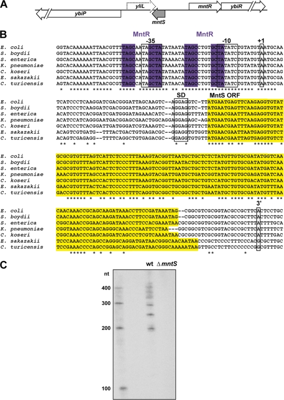
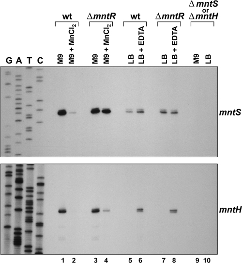
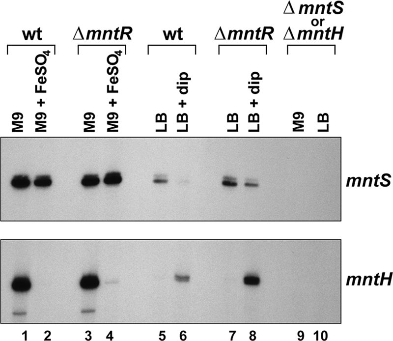
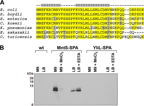
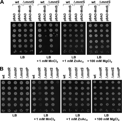
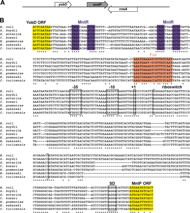
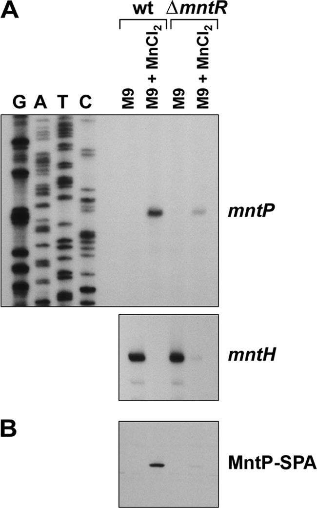
Manganese is a critical micronutrient for cells, serving as an enzyme cofactor and protecting against oxidative stress. Yet, manganese is toxic in excess and little is known about its distribution in cells. Bacteria control intracellular manganese levels by the transcription regulator MntR. When this work began, the only Escherichia coli K-12 gene known to respond to manganese via MntR repression was mntH, which encodes a manganese importer. We show that mntS (formerly the small RNA gene rybA) is repressed by manganese through MntR and encodes an unannotated 42-amino-acid protein. Overproduction of MntS causes manganese sensitivity, while a lack of MntS perturbs proper manganese-dependent repression of mntH. We also provide evidence that mntP (formerly yebN), which encodes a putative efflux pump, is positively regulated by MntR. Deletion of mntP leads to profound manganese sensitivity and to elevated intracellular manganese levels. This work thus defines two new proteins involved in manganese homeostasis and suggests mechanisms for their action.


Figures

Fig. 1.
Fig. 1.
The mntS locus, sequence alignment, and transcript profile. (A) Schematic of the mntS genomic locus. (B) Alignment of the mntS gene, showing the consensus MntR binding motif (purple), predicted −10 and −35 promoter regions, mapped transcription start site (+1) at position 852270 in the MG1655 genome, consensus Shine-Dalgarno (SD) sequence, MntS open reading frame (ORF; yellow), and the first 3′ end of the mntS transcript at position 852065. Species included Escherichia coli, Shigella boydii, Salmonella enterica, Citrobacter koseri, Klebsiella pneumoniae, Enterobacter sakazakii, and Cronobacter turicensis. (C) Northern blot analysis of the mntS transcript from wild-type (MG1655) and ΔmntS (GSO459) cells grown to mid-exponential phase in M9-glucose medium.
Fig. 2.
Fig. 2.
Expression of mntS and mntH in response to divalent cations. Primer extension analysis of the mntS and mntH transcripts from wild-type (wt; MG1655), ΔmntR (GSO458), ΔmntS (GSO459) (top), and ΔmntH (GSO460) (bottom) cultures during growth to mid-exponential phase in M9-glucose or LB after mock treatment or exposure to 10 μM MnCl2 or 1 mM EDTA, respectively, for 30 min. Note that the sequencing ladder corresponds to the strand complementary to the open reading frames.
Fig. 3.
Fig. 3.
Expression of mntS and mntH in response to iron. Primer extension analysis of the mntS and mntH transcripts from wild-type (wt; MG1655), ΔmntR (GSO458), ΔmntS (GSO459) (top), and ΔmntH (GSO460) (bottom) cultures during growth to mid-exponential phase in M9-glucose or LB after mock treatment or exposure to 10 μM FeSO4 or 0.5 mM 2,2′-dipyridyl (dip), respectively, for 30 min.
Fig. 4.
Fig. 4.
Conservation and expression of the MntS small protein. (A) Alignment of the 42-amino-acid MntS open reading frame. Strictly conserved residues are highlighted in yellow, and similar residues are shown in gray. Predicted α-helical regions are indicated above by the letter H. Genus abbreviations are as for Fig. 1. (B) Western blot analysis of wild-type (wt; MG1655), MntS-SPA (GSO462), and YliL-SPA (GSO463) cells grown to mid-exponential phase in M9-glucose or LB and mock treated or exposed to 10 μM MnCl2 or 1 mM EDTA, respectively, for 2 h. The expected molecular masses of the proteins were as follows: MntS-SPA, ∼13 kDa; YliL-SPA, ∼18 kDa; SPA tag, ∼8 kDa.
Fig. 5.
Fig. 5.
Metal sensitivity associated with MntS overproduction or a lack of MntP. (A) Ten-fold serial dilutions of mid-exponential-phase cultures of wild-type (wt; MG1655) or ΔmntS (GSO459) cells bearing the pBAD24 empty vector or pBAD24-MntS were spotted onto LB plates containing 0.2% arabinose, 100 μg/ml ampicillin, and the indicated amounts of metals. The effects of 4 mM FeSO4, 6 mM FeCl3, 2 mM NiCl2, 3 mM CuCl2, 1 mM CoCl2, and 1 mM CdSO4 were similarly tested (data not shown). (B) Ten-fold serial dilutions of mid-exponential-phase cultures of wild-type (MG1655), ΔmntR (GSO458), ΔmntS (GSO459), ΔmntH (GSO460), and ΔmntP (GSO461) were spotted onto LB plates containing the indicated amounts of metals. The effects of 4 mM FeSO4, 6 mM FeCl3, 2 mM NiCl2, and 3 mM CuCl2 were similarly tested (data not shown).
Fig. 6.
Fig. 6.
Lack of mntH repression in the ΔmntS strain. Primer extension analysis monitored the mntH and mntS transcript levels, and Western blot analysis was used to determine MntS-SPA expression in stationary-phase cultures of wild-type (wt; MG1655) or ΔmntS (GSO459) strains grown overnight in M9-glucose medium containing the indicated concentration of MnCl2.
Fig. 7.
Fig. 7.
The mntP locus and sequence alignment. (A) Schematic of the mntP genomic locus. (B) Alignment of the mntP promoter and 5′-untranslated region, showing the end of the upstream YobD open reading frame (ORF; yellow), consensus MntR (purple), and Fur (orange) binding motifs, predicted −10 and −35 regions, approximate transcription start site (+1) mapped to within ∼5 nt (MG1655 coordinates 1903482 to 1903487), Shine-Dalgarno (SD) sequence, and the beginning of the MntP open reading frame (yellow). Genus abbreviations are as for Fig. 1.
Fig. 8.
Fig. 8.
Expression of mntP in response to manganese. (A) Primer extension analysis of the mntP and mntH transcripts from wild-type (wt; MG1655) and ΔmntR (GSO458) cultures during growth to mid-exponential phase in M9-glucose after mock treatment or exposure to 10 μM MnCl2 for 10 min. (B) Western blot analysis of MntP-SPA expression levels from MntP-SPA (GSO464) or ΔmntR MntP-SPA (GSO465) cultures grown as described for panel A.

References

    1. Anjem A., Varghese S., Imlay J. A. 2009. Manganese import is a key element of the OxyR response to hydrogen peroxide in Escherichia coli. Mol. Microbiol. 72:844–858 - PMC - PubMed
    1. Barrick J. E., et al. 2004. New RNA motifs suggest an expanded scope for riboswitches in bacterial genetic control. Proc. Natl. Acad. Sci. U. S. A. 101:6421–6426 - PMC - PubMed
    1. Cherepanov P. P., Wackernagel W. 1995. Gene disruption in Escherichia coli: TcR and KmR cassettes with the option of Flp-catalyzed excision of the antibiotic-resistance determinant. Gene 158:9–14 - PubMed
    1. Cotruvo J. A., Stubbe J. 2011. Escherichia coli class Ib ribonucleotide reductase contains a dimanganese(III)-tyrosyl radical cofactor in vivo. Biochemistry 50:1672–1681 - PMC - PubMed
    1. Cromie M. J., Shi Y., Latifi T., Groisman E. A. 2006. An RNA sensor for intracellular Mg2+. Cell 125:71–84 - PubMed

Publication types

MeSH terms