Comparative Study
. 2011 Dec;80(6):1076-84.
doi: 10.1124/mol.111.073585. Epub 2011 Sep 9.

Restoring soluble guanylyl cyclase expression and function blocks the aggressive course of glioma

Affiliations
Comparative Study

Restoring soluble guanylyl cyclase expression and function blocks the aggressive course of glioma

Haifeng Zhu et al. Mol Pharmacol. 2011 Dec.

Abstract

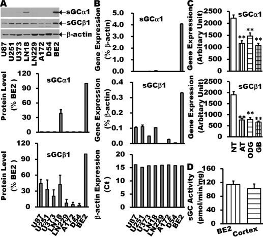
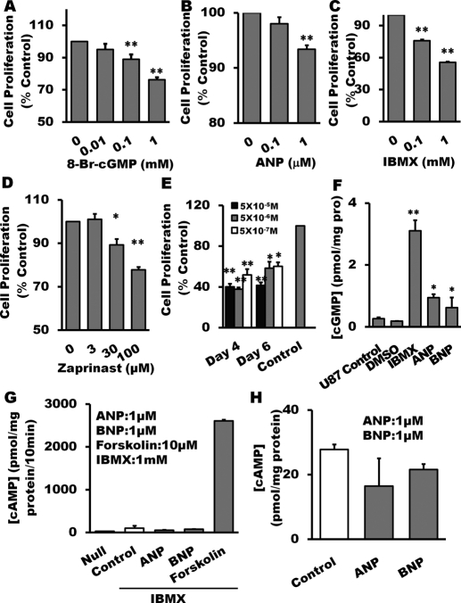
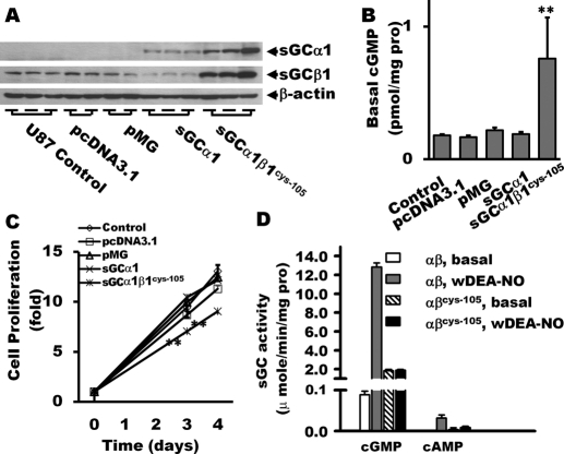
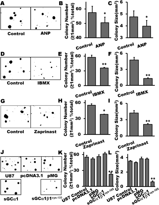
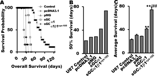
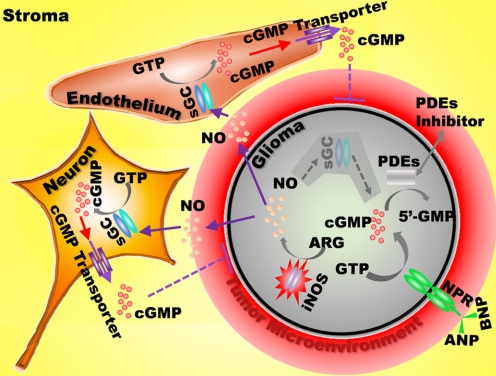
The NO and cGMP signaling pathways are of broad physiological and pathological significance. We compared the NO/soluble guanylyl cyclase (sGC)/cGMP pathway in human glioma tissues and cell lines with that of healthy control samples and demonstrated that sGC expression is significantly lower in glioma preparations. Our analysis of GEO databases (National Cancer Institute) further revealed a statistically significant reduction of sGC transcript levels in human glioma specimens. On the other hand, the expression levels of particulate (membrane) guanylyl cyclases (pGC) and cGMP-specific phosphodiesterase (PDE) were intact in the glioma cells that we have tested. Pharmacologically manipulating endogenous cGMP generation in glioma cells through either stimulating pGC by ANP/BNP, or blocking PDE by 3-isobutyl-1-methylxanthine/zaprinast caused significant inhibition of proliferation and colony formation of glioma cells. Genetically restoring sGC expression also correlated inversely with glioma cells growth. Orthotopic implantation of glioma cells transfected with an active mutant form of sGC (sGCα1β1(Cys105)) in athymic mice increased the survival time by 4-fold over the control. Histological analysis of xenografts overexpressing α1β1(Cys105) sGC revealed changes in cellular architecture that resemble the morphology of normal cells. In addition, a decrease in angiogenesis contributed to glioma inhibition by sGC/cGMP therapy. Our study proposes the new concept that suppressed expression of sGC, a key enzyme in the NO/cGMP pathway, may be associated with an aggressive course of glioma. The sGC/cGMP signaling-targeted therapy may be a favorable alternative to chemotherapy and radiotherapy for glioma and perhaps other tumors.


Figures

Fig. 1.
Fig. 1.
Reduction of sGC expression in human glioma tissues and cell lines. sGC expression in glioma cell lines (U87, U251, U373, A172, LN18, LN229, and D54) was examined by Western blot (A) and real time-Q-PCR (B) and compared with that in BE2 human neuroblastoma cell line, which normally expresses both sGC α1 and β1 subunits at levels similar to those in normal human cortex (D) (Bonkale et al., 1995; Corbalán et al., 2002; Sharina et al., 2008). The GEO database analysis of sGC gene expression in human glioma tissues of different grade (C) showed that the expression of sGC α1 and β1 is markedly decreased in astrocytoma (n = 26), oligodendrocytoma (n = 50), and glioblastoma multiforme (n = 81) compared with normal brain tissues (n = 23). Data are mean ± S.E.M. n = 3 to 6 per group for glioma cell lines. **, P < 0.01 (versus normal brain tissues).
Fig. 2.
Fig. 2.
pGCs, PDEs, and PKG expression in glioma cell lines. Real time-Q-PCR analysis of glioma cell lines (U87, U251, U373, A172, LN18, LN229, D54) determined expression profiles of NPR1, -2, -3 (A), PDEs (B), and PKG (D and E). The profile of PDE isoform in U87 cells was also assayed (C). Data are mean ± S.E.M. n = 3 to 6 per group for glioma cell lines.
Fig. 3.
Fig. 3.
Inhibition of glioma cell proliferation by pharmacological restoration of cGMP. Treatment with 8-bromo-cGMP (A), particulate GC agonist ANP (B), nonselective PDE inhibitor IBMX (C), and PDE5-specific inhibitor zaprinast (D) for 24 h, respectively, suppressed U87 cell proliferation measured by MTT assay. Combined treatment of ANP with 0.1 mM IBMX for 4 or 6 days inhibited U87 cell proliferation (E). After 24-h treatment with ANP (1 μM), IBMX (1 mM), and 8-Br-cAMP (1 mM), cGMP accumulation in U87 glioma cells was increased significantly (F). Adenylyl cyclase activity (G) and cAMP accumulation (H) in U87 glioma cells was measured after stimulation with cGMP-promoting reagents. 10 μM forskolin was used as positive control. Data are mean ± S.E.M. n = 6 per group, *, P < 0.05; **, P < 0.01 (versus control groups).
Fig. 4.
Fig. 4.
Proliferation analysis of sGCα1 and sGC α1β1Cys105-expressing stable clones. Overexpression of sGCα1 and sGCα1β1Cys105 mutant in U87 glioma cells was confirmed by Western blot (A). Lanes 1 to 3, U87 control; lanes 4 to 7, vector alone; lanes 8 and 9, sGCα1 overexpression; and lanes 11 to 13, sGCα1β1Cys105 overexpression. The lysates were also probed for the basal cGMP levels in U87 glioma cells (B). Codelivery of sGCα1 and sGCβ1Cys105 in U87 glioma cells suppressed cellular proliferation (C). Basal and NO-stimulated activity (1 μM DEA-NO) of the wild-type and mutant αβCys105 sGC in generation of cGMP and cAMP were evaluated (D). n = 6 per group for cGMP and cAMP assays; n = 18 per group for proliferation assay (MTT). **, P < 0.01 (versus control groups).
Fig. 5.
Fig. 5.
Inhibition of glioma colony formation by pharmacological restoration of cGMP or genetical restoration of sGC. Treatment with 1 μM ANP (A–C), IBMX (0.1 μM; D–F) and zaprinast (30 μM, G–I), respectively, markedly inhibited the number of larger colonies with the size ≥1 mm2 and reduced average size of colonies of U87 glioma cells. The clones of the U87 glioma cell with stable overexpression of α1β1Cys105 sGC formed fewer and smaller colonies (J–L). Reagents were freshly added every 3 days for a period of 3 weeks. Colonies were counted at day 21 using an inverted microscope with a digital camera photo system. Data are mean ± S.E.M. n = 6 per group, *, P < 0.05; **, P < 0.01 (versus control groups).
Fig. 6.
Fig. 6.
Intracerebral human glioma xenograft assay for sGC transfectants. In vivo antitumor activity of sGC / cGMP was evaluated in athymic mice with intracerebral xenotransplantation of U87 cells with or without transfection. The survival probability (A), average survival time (B), and 50% survival time (C) were plotted. Data are mean ± S.E.M. n = 6 for U-87 control, U87 with empty vectors, and U87-overexpressing sGCα1; n = 18 for U87 glioma cells transfected with α1β1Cys105 sGC subunits. **, P < 0.01(versus control groups). ##, P < 0.01 (versus sGCα1-overexpressing group).
Fig. 7.
Fig. 7.
Characterization of glioma xenograft with sGC transfectants. H.E. staining showed tumor mass derived from sGC α1β1Cys105-overexpressing cells with well defined pathological features of glioblastoma (A; 100× and 400× magnification). The xenografts with α1β1Cys105 sGC are composed of heterogeneous populations of tumor cells (A, 400× magnification) with different nuclear density. The tumor tissue derived from α1β1Cys105 sGC transfected clone had significantly fewer microvessels according to CD31 staining (B and C; 200× magnification). Significant reduction in Ki-67 staining, a marker associated with cell proliferation, was observed in sGC transfectant-derived tumors (D and E; 200× magnification). Data are mean ± S.E.M. n = 3 to 4 for each group. **, P < 0.01(versus control groups).
Scheme 1.
Scheme 1.
NO/cGMP signaling in malignant tumor. NO and cGMP participate in regulation of inflammation in the cancer microenvironment and contribute to proliferation and/or differentiation of tumor cells and surrounding supporting cells. The iNOS/NO/cGMP signaling pathway is not functional in glioma because of sGC-deficiency. However, higher concentration of NO generated by malignant cells and/or its microenvironment can penetrate cell membrane and increase cGMP levels in nonmalignant stroma cells. cGMP can be released into the intercellular space through cGMP transporters; however, extracellular cGMP cannot cross the cell membrane to re-enter the cells.

References

    1. Basson MD, Hong F. (1996) Regulation of human Caco-2 intestinal epithelial brush border enzyme activity by cyclic nucleotides. Cancer Lett 99:155–160 - PubMed
    1. Bhuiyan MB, Murad F, Fant ME. (2006) The placental cholinergic system: localization to the cytotrophoblast and modulation of nitric oxide. Cell Commun Signal 4:4. - PMC - PubMed
    1. Bian K, Doursout MF, Murad F. (2008) Vascular system: role of nitric oxide in cardiovascular diseases. J Clin Hypertens (Greenwich) 10:304–310 - PMC - PubMed
    1. Bian K, Gao Z, Weisbrodt N, Murad F. (2003) The nature of heme/iron-induced protein tyrosine nitration. Proc Natl Acad Sci USA 100:5712–5717 - PMC - PubMed
    1. Bian K, Harari Y, Zhong M, Lai M, Castro G, Weisbrodt N, Murad F. (2001) Down-regulation of inducible nitric-oxide synthase (NOS-2) during parasite-induced gut inflammation: a path to identify a selective NOS-2 inhibitor. Mol Pharmacol 59:939–947 - PubMed

Publication types

MeSH terms

Substances

LinkOut - more resources