Review
. 2012 Jun;18(3):216-23.
doi: 10.1177/1073858411418525. Epub 2011 Sep 9.

Presynaptically silent synapses: dormancy and awakening of presynaptic vesicle release

Affiliations
Review

Presynaptically silent synapses: dormancy and awakening of presynaptic vesicle release

Devon C Crawford et al. Neuroscientist. 2012 Jun.

Abstract

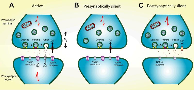
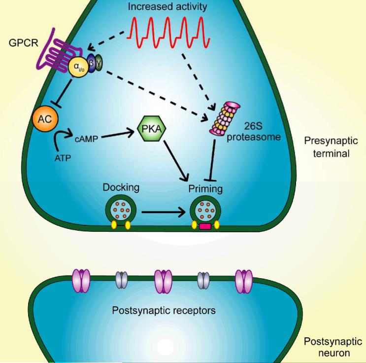
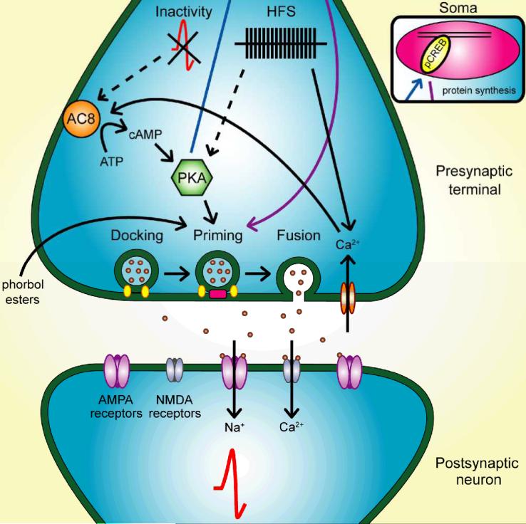
Synapses represent the main junctures of communication between neurons in the nervous system. In many neurotransmitter systems, a fraction of presynaptic terminals fails to release vesicles in response to action potential stimulation and strong calcium influx. These silent presynaptic terminals exhibit a reversible functional dormancy beyond low vesicle release probability, and dormancy status may have important implications in neural function. Recent advances have implicated presynaptic proteins interacting with vesicles downstream of cAMP and protein kinase A signaling cascades in modulating the number of these mute presynaptic terminals, and dormancy induction may represent a homeostatic neuroprotective mechanism active during pathological insults involving excitotoxicity. Interestingly, dormancy reversal may also be induced during Hebbian plasticity. Here, details of synaptic dormancy, recent insights into the molecular signaling cascades involved, and potential clinical and mechanistic implications of this form of synaptic plasticity are described.


Figures

Figure 1
Figure 1
Categories of silent synapses. A. Active synapses consist of presynaptic terminals with functional vesicle docking, priming, and release upon calcium influx, all powered primarily by mitochondrial ATP production. The probability of vesicle release (pr) is modulated without altering the qualitative release competence of the terminal. Neurotransmitter released from active presynaptic terminals binds to postsynaptic receptors and causes a postsynaptic response. At glutamate synapses, for example, glutamate released via presynaptic vesicle fusion will bind to AMPA receptors, allowing net cation influx that directly depolarizes the target cell and relieves voltage-dependent magnesium block of NMDA receptors (not depicted). Depolarizing effects of activated AMPA and NMDA receptors contribute to action potential generation. B. In presynaptically silent synapses, vesicle docking is intact, but priming and fusion are impaired, even with strong depolarization and calcium influx that overcomes low vesicle release probability. Without transmitter release, there is no postsynaptic response. C. Postsynaptically silent synapses maintain active presynaptic terminals, but the postsynaptic membrane is missing receptors necessary to generate a response. At glutamate synapses, AMPA receptors are absent, leaving NMDA receptors unable to overcome voltage-dependent block.
Figure 2
Figure 2
Malleability in the number of dormant presynaptic terminals. A. The percentage of active glutamate terminals is measured using FM1-43 dye uptake (green) and its co-localization with a presynaptic marker (vesicular glutamate transporter 1 or vGluT-1; red); dormant terminals are, therefore, represented by red puncta without any overlying green. Activity also modulates the number of dormant terminals. For example, co-localization occurs more often in control than in depolarized (4 hr 30 mM KCl) cultured hippocampal neurons. This suggests that fewer glutamatergic presynaptic terminals are competent to recycle vesicles after depolarization. Modified with permission from Crawford, Chang, Hyrc, and Mennerick 2011. B. The percentage of active terminals correlates with the size of the readily releasable pool of vesicles. Excitatory postsynaptic currents (EPSCs) measured in autaptic cultured hippocampal neurons are depressed after prolonged (16 hr) depolarization with 30 mM KCl. Top: EPSCs were elicited after action potential stimulation, which are probabilistically dependent on calcium influx into the presynaptic terminal. Bottom: EPSCs were elicited by application of hypertonic sucrose, which causes calcium-independent fusion of all release-ready vesicles. Because both types of EPSCs are depressed after depolarization, this suggests that the size of the readily releasable pool of vesicles is decreased rather than the probability of vesicle release. C. The percentage of glutamatergic (red) terminals in cultured hippocampal neurons that take up the dye FM1-43 (green) increases after 4 hr 50 μM forskolin treatment, which increases adenylyl cyclase activity and, therefore, cAMP production. This suggests that more terminals are release-competent after increased cAMP signaling. Modified with permission from Moulder and others 2008. D. EPSCs in autaptic hippocampal neurons are increased after 4 hr 50 μM forskolin application. Because both calcium-dependent action-potential evoked EPSCs and calcium-independent sucrose-evoked EPSCs are increased after forskolin, this suggests that the readily releasable pool is increased rather than the probability of vesicle release. Modified with permission from Moulder and others 2008.
Figure 3
Figure 3
Signaling cascades participating in presynaptic dormancy induction. Prolonged strong depolarization or increased action potential firing induces presynaptic dormancy through activation of inhibitory G-proteins and through activation of the ubiquitin-proteasome system. Depolarization increases proteasome activity through unknown mechanisms. Both depolarization- and G-protein-coupled receptor (GPCR) agonist-induced silencing require proteasome activity. Dormancy is also induced via reduced cAMP signaling, so inhibitory actions of the Gα subunit on adenylyl cyclase (AC) likely reduce cAMP and protein kinase A (PKA) signaling during silencing induction. PKA phosphorylates presynaptic priming proteins like Rim1, a modification that may render Rim1 resistant to proteasome degradation; therefore, less Rim1 phosphorylation is expected after depolarization. Increased proteasome activity, combined with a vulnerable presynaptic protein population, may then lead to priming protein degradation. This model provides a plausible mechanism for priming protein level reduction and dormancy induction by depolarization. Postsynaptic protein levels are unaltered by induction of presynaptic dormancy.
Figure 4
Figure 4
Signaling cascades participating in presynaptic dormancy reduction. Under some conditions, inactivity and increased cAMP activate previously dormant terminals. Calcium-dependent adenylyl cyclase VIII (AC8) knockout prevents recovery of active terminals when elevated neuronal activity is removed, so AC8 may be the cAMP source responsible for this form of unsilencing. In other contexts multiple bursts of high frequency stimulation (HFS), which often causes strong calcium influx and long-term potentiation, lead to presynaptic awakening. Both activity reduction and HFS require protein kinase A (PKA) signaling for presynaptic activation, but how ostensibly opposite changes in activity both recruit PKA in different experimental contexts remains unknown. Once PKA is activated, phosphorylation events may slow priming protein degradation. Additionally, PKA phosphorylates and thereby activates nuclear transcription factors (blue arrow and inset) like cAMP response element-binding protein (CREB), and this may increase synthesis of presynaptic proteins vital for vesicle priming and release (purple arrow and inset). A third pathway to unsilencing is via phorbol esters, which enhance function of the priming protein Munc13-1. Under this model of dormancy reduction, postsynaptic responses are restored once the presynaptic terminal regains the ability to release neurotransmitter.

References

    1. Alberini CM, Ghirardi M, Huang YY, Nguyen PV, Kandel ER. A molecular switch for the consolidation of long-term memory: cAMP-inducible gene expression. Ann N Y Acad Sci. 1995;758:261–86. - PubMed
    1. Altrock WD, tom Dieck S, Sokolov M, Meyer AC, Sigler A, Brakebusch C. Functional inactivation of a fraction of excitatory synapses in mice deficient for the active zone protein bassoon. Neuron. 2003;37(5):787–800. others. - PubMed
    1. Augustin I, Rosenmund C, Sudhof TC, Brose N. Munc13-1 is essential for fusion competence of glutamatergic synaptic vesicles. Nature. 1999;400(6743):457–61. - PubMed
    1. Betz A, Ashery U, Rickmann M, Augustin I, Neher E, Sudhof TC. Munc13-1 is a presynaptic phorbol ester receptor that enhances neurotransmitter release. Neuron. 1998;21(1):123–36. others. - PubMed
    1. Bolshakov VY, Golan H, Kandel ER, Siegelbaum SA. Recruitment of new sites of synaptic transmission during the cAMP-dependent late phase of LTP at CA3-CA1 synapses in the hippocampus. Neuron. 1997;19(3):635–51. - PubMed

Publication types

MeSH terms

LinkOut - more resources