. 2011 Nov;115(5):992-1002.
doi: 10.1097/ALN.0b013e3182303a63.

General Anesthesia Causes Long-term Impairment of Mitochondrial Morphogenesis and Synaptic Transmission in Developing Rat Brain

Affiliations

General Anesthesia Causes Long-term Impairment of Mitochondrial Morphogenesis and Synaptic Transmission in Developing Rat Brain

Victoria Sanchez et al. Anesthesiology. 2011 Nov.

Abstract

Background: Clinically used general anesthetics, alone or in combination, are damaging to the developing mammalian brain. In addition to causing widespread apoptotic neurodegeneration in vulnerable brain regions, exposure to general anesthesia at the peak of synaptogenesis causes learning and memory deficiencies later in life. In vivo rodent studies have suggested that activation of the intrinsic (mitochondria-dependent) apoptotic pathway is the earliest warning sign of neuronal damage, suggesting that a disturbance in mitochondrial integrity and function could be the earliest triggering events.

Methods: Because proper and timely mitochondrial morphogenesis is critical for brain development, the authors examined the long-term effects of a commonly used anesthesia combination (isoflurane, nitrous oxide, and midazolam) on the regional distribution, ultrastructural properties, and electron transport chain function of mitochondria, as well as synaptic neurotransmission, in the subiculum of rat pups.

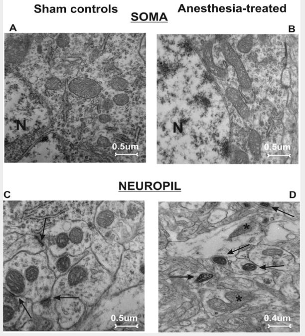
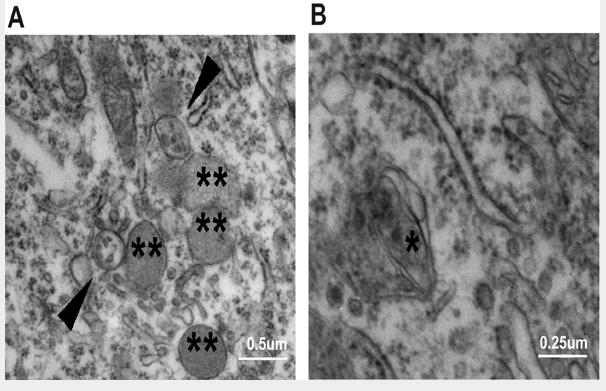
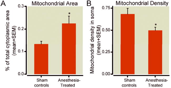
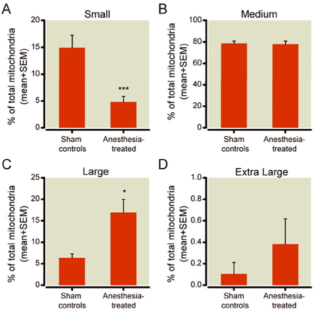
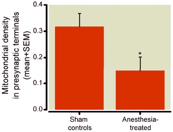
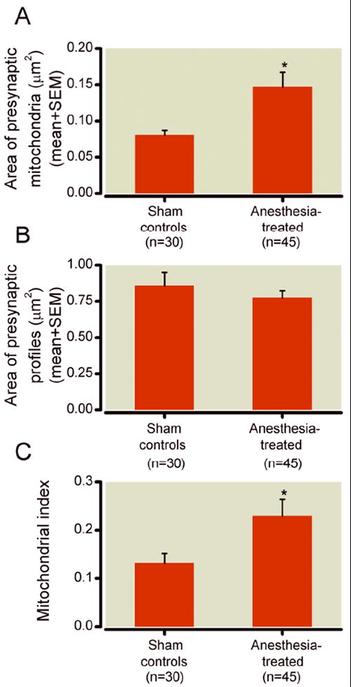
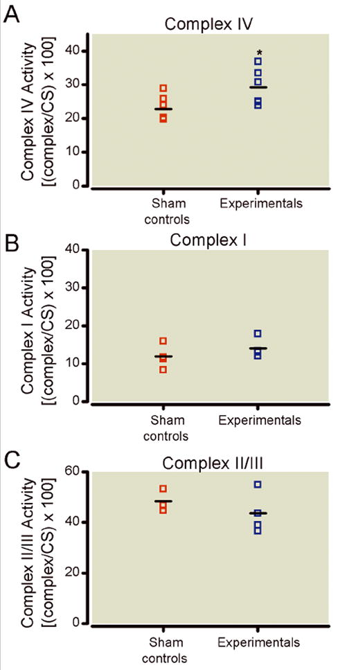
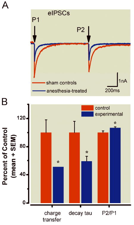
Results: This anesthesia, administered at the peak of synaptogenesis, causes protracted injury to mitochondria, including significant enlargement of mitochondria (more than 30%, P < 0.05), impairment of their structural integrity, an approximately 28% increase in their complex IV activity (P < 0.05), and a twofold decrease in their regional distribution in presynaptic neuronal profiles (P < 0.05), where their presence is important for the normal development and functioning of synapses. Consequently, the authors showed that impaired mitochondrial morphogenesis is accompanied by heightened autophagic activity, decrease in mitochondrial density (approximately 27%, P < 0.05), and long-lasting disturbances in inhibitory synaptic neurotransmission. The interrelation of these phenomena remains to be established.

Conclusion: Developing mitochondria are exquisitely vulnerable to general anesthesia and may be important early target of anesthesia-induced developmental neurodegeneration.


Figures

Figure 1
Figure 1. Anesthesia causes long-lasting ultrastructural changes in mitochondria in subiculi of 21-day-old rats
A, C) The pyramidal neuron (A) and neuropil (C; synaptic contacts are noted with arrows) in a subiculum from a control rat show abundant small mitochondria with no evidence of swelling or injury. B, D) Mitochondria in the perikarion of a pyramidal neuron (B) and nerve terminals in a neuropil (D) of subiculum from experimental rats display structural disorganization of cristae (asterisks), as well as dilated intracristal spaces with vacuoles and overall swelling. Note the presence of dark, condensed mitochondria in late stages of degeneration (arrows) (magnification 12,000×). N - nucleus.
Figure 2
Figure 2. Anesthesia promotes autophagic activity, as shown in subicular pyramidal neurons of 21-day-old rats
A) In experimental pyramidal neurons, numerous lysosomes (double asterisks) and autophagic vacuoles (arrowheads) were dispersed throughout the cytoplasm. B) Autophagosomes, double-layered membrane structures, were frequently noted in experimental neurons where parts of cannibalized mitochondria could be detected (single asterisk).
Figure 3
Figure 3. Morphometric analysis of mitochondria in the perikaryon of pyramidal subicular neurons of 21-day-old rats
A) Mitochondria in the experimental neurons occupy significantly more cytoplasmic soma than do those in controls (22.5% vs. 13.44%,* p < 0.05) (n = 15 neurons per group from 3 animals each). B) In experimental animals, mitochondrial density, presented as the number of mitochondria per unit area (μm2) of cytoplasmic soma, is significantly lower in pyramidal neurons than that in controls (* p < 0.05, n = 15 neurons per group from 3 control and 3 experimental pups; control and experimental pups were litter-matched).
Figure 4
Figure 4. Mitochondrial size classification in the perikaryon of pyramidal subicular neurons of 21-day-old rats
A) A small fraction of mitochondria (up to 0.05 μm2) constitutes about 15% of total mitochondria in control pyramidal neurons, but only 5% in the experimental pyramidal neurons (*** p < 0.001). B) A medium-sized fraction of mitochondria (0.06 to 0.25 μm2) represents the largest population of mitochondria in subicular pyramidal neurons. This fraction remains unchanged after anesthesia treatment. C) A large fraction of mitochondria (0.26 to 0.65 μm2) constitutes only about 5% of total mitochondria in control pyramidal neurons, but 15% of those in experimental pyramidal neurons (* p < 0.05). D) An extra-large fraction of mitochondria (more than 0.65 μm2) represents less than 1% of the total number of mitochondria in experimental pyramidal neurons and shows over two-fold higher prevalence in these neurons (fig. 3D) although it did not reach statistical significance (n = 15 neurons per group from 3 control and 3 experimental pups; control and experimental pups were litter-matched).
Figure 5
Figure 5. Anesthesia decreases mitochondrial density in presynaptic neuronal terminal subicular neuropils of 21-day-old rats
Compared to controls, fewer presynaptic neuronal terminals in experimental subicular neuropils contain mitochondrial profiles. When the findings are presented as a percentage of presynaptic profiles containing mitochondria, a significantly (about two-fold) higher percentage of mitochondria-containing presynaptic profiles occurs in control subiculi than in experimental subiculi (* p < 0.05) (n = 28 photo frames/group from 4 control and 4 experimental pups from two different litters; control and experimental pups were litter-matched).
Figure 6
Figure 6. Morphometric analysis of mitochondria in presynaptic neuronal terminals in subicular neuropils of 21-day-old rats
A) Morphometric analysis of the area of mitochondrial profiles shows that experimental mitochondrial profiles were approximately 30% larger than that of controls (* p < 0.05)) (n = 30 photo frames obtained from 5 control pups; n = 45 photo frames obtained from 5 experimental pups; control and experimental pups were litter-matched; total of three different litters were used). B) There is no difference between control and experimental subiculi with regard to the areas of mitochondria-containing presynaptic nerve terminals (n = 30 photo frames obtained from 5 control pups; n = 45 photo frames obtained from 5 experimental pups; control and experimental pups were litter-matched; total of three different litters were used). C) The calculated mitochondrial index (ratio between mitochondrial area and mitochondria-containing presynaptic area) was significantly higher in experimental subicular neuropils (* p < 0.05) than in control neuropils.
Figure 7
Figure 7
Anesthesia differentially modulates the activity of mitochondrial respiratory chain proteins. A) Compared to control subiculi, the activity of complex IV in experimental subiculi is significantly increased at 24 h after anesthesia (* p < 0.05) (n = 8 pups in control group; n = 6 pups in experimental group). B) The activity of complex I was unchanged in the anesthesia-treated group as compared to sham controls (n = 5 pups per group). C) The activity of complex II was unchanged in the anesthesia-treated group as compared to sham controls (n = 3 pups in control group; n = 4 pups in experimental group). The activity of complexes I, II, and IV were expressed as ratios [per the activity of citrate synthase] since citrate synthase activity is directly proportional to mitochondrial content.
Figure 8
Figure 8. Alterations occurred in inhibitory synaptic transmission in pyramidal cells of subiculi after exposure to anesthesia early in life
A) Representative eIPSCs obtained using a paired-pulse protocol to record from two pyramidal cells in the subiculi of rats in the control (red trace) and experimental groups (blue trace). Note that the experimental group had decreased current amplitude and faster decay. Arrows indicate the time of paired-pulse stimulus application (interval 1.1 s). Stimulus transients have been removed for clarity of the current traces. B) Histograms showing average data from control cells (n = 14) and experimental cells (n = 12). Black solid bars indicate control cells; gray bars represent experimental cells; vertical lines indicate the SEM of multiple determinations. All data are normalized to 100% of average responses in the control group. Left panel shows a decrease in net charge transfer of eIPSCs from 100 ± 18% to 51 ± 9% (p < 0.05) in the experimental group; middle panels show a decrease in decay of tau from 100 ± 16% to 59 ± 7%; right panels show a small but significant increase in P2/P1 from 100 ± 2% to 107 ± 1% (p < 0.05).

Similar articles

Cited by

References

    1. Yon J-H, Daniel-Johnson J, Carter LB, Jevtovic-Todorovic V. Anesthesia induces neuronal cell death in the developing rat brain via the intrinsic and extrinsic apoptotic pathways. Neuroscience. 2005;35:815–27. - PubMed
    1. Wilder RT, Flick RP, Sprung J, Katusic SK, Barbaresi WJ, Mickelson C, Gleich SJ, Schroeder DR, Weaver AL, Warner DO. Early exposure to anesthesia and learning disabilities in a population-based birth cohort. Anesthesiology. 2009;110:796–804. - PMC - PubMed
    1. Yon J-H, Carter LB, Reiter RJ, Jevtovic-Todorovic V. Melatonin reduces the severity of anesthesia-induced apoptotic neurodegeneration in the developing rat brain. Neurobiol Dis. 2006;21:522–30. - PubMed
    1. Head BP, Patel HH, Niesman IR, Drummond JC, Roth DM, Patel PM. Inhibition of p75 neurotrophin receptor attenuates isoflurane-mediated neuronal apoptosis in the neonatal central nervous system. Anesthesiology. 2009;110:813–25. - PMC - PubMed
    1. Lunardi N, Ori C, Erisir A, Jevtovic-Todorovic V. General anesthesia causes long-lasting disturbances in the ultrastructural properties of developing synapses in young rats. Neurotox Res. 2010;17:179–88. - PMC - PubMed

Publication types

Substances