. 2011 Nov 16;30(22):4571-85.
doi: 10.1038/emboj.2011.325.

IRF6 is a mediator of Notch pro-differentiation and tumour suppressive function in keratinocytes

Affiliations

IRF6 is a mediator of Notch pro-differentiation and tumour suppressive function in keratinocytes

Gaetana Restivo et al. EMBO J. .

Abstract

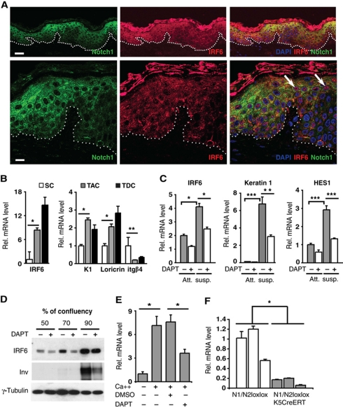
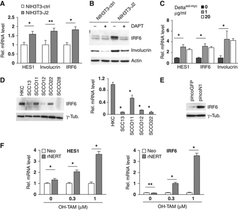
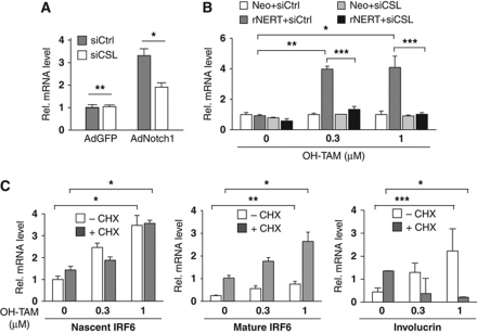
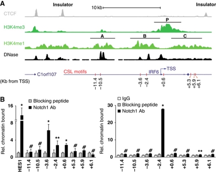
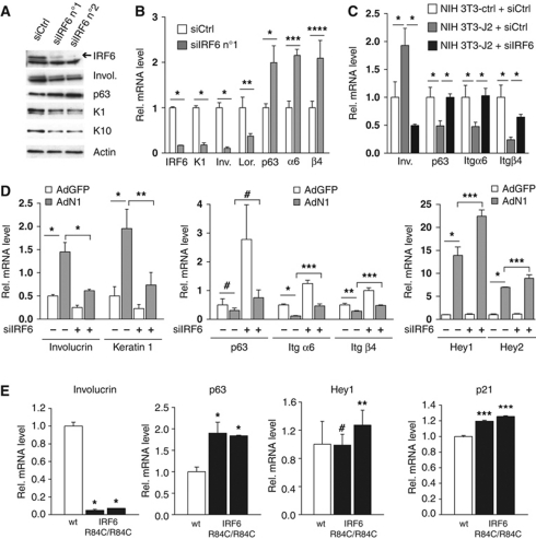
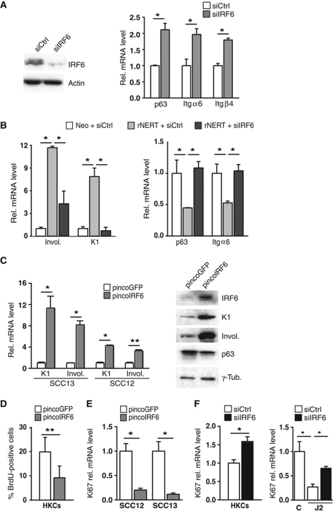
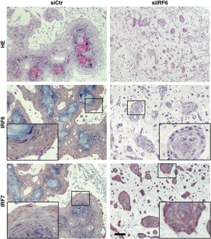
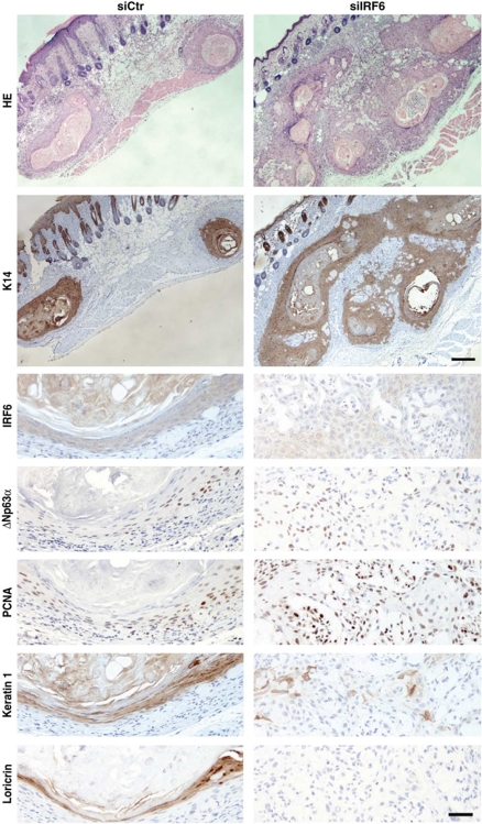
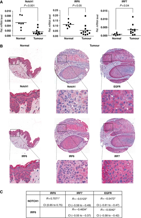
While the pro-differentiation and tumour suppressive functions of Notch signalling in keratinocytes are well established, the underlying mechanisms remain poorly understood. We report here that interferon regulatory factor 6 (IRF6), an IRF family member with an essential role in epidermal development, is induced in differentiation through a Notch-dependent mechanism and is a primary Notch target in keratinocytes and keratinocyte-derived SCC cells. Increased IRF6 expression contributes to the impact of Notch activation on growth/differentiation-related genes, while it is not required for induction of 'canonical' Notch targets like p21(WAF1/Cip1), Hes1 and Hey1. Down-modulation of IRF6 counteracts differentiation of primary human keratinocytes in vitro and in vivo, promoting ras-induced tumour formation. The clinical relevance of these findings is illustrated by the strikingly opposite pattern of expression of Notch1 and IRF6 versus epidermal growth factor receptor in a cohort of clinical SCCs, as a function of their grade of differentiation. Thus, IRF6 is a primary Notch target in keratinocytes, which contributes to the role of this pathway in differentiation and tumour suppression.


Conflict of interest statement

The authors declare that they have no conflict of interest.

Figures

Figure 1
Figure 1
IRF6 is induced during keratinocyte differentiation through a Notch-dependent mechanism. (A) Confocal double-immunofluorescence analysis of Notch1 and IRF6 expression in human skin. (Upper panels) Representative low-magnification images showing concomitantly increased expression of the proteins in the suprabasal epidermal layers. (Lower panels) High-magnification images showing prevalent cytoplasmic localization of the two proteins, with nuclear localization of IRF6 being also detectable in some cells of the outer layers (indicated with arrows). The prevalent cytoplasmic staining of the two proteins is consistent with previous publications (Rangarajan et al, 2001; Ingraham et al, 2006; Richardson et al, 2006). Images are representative of several independent fields. The IRF6 results were confirmed by additional immunofluorescence/confocal imaging analysis of human skin (Supplementary Figure S1). Bars=30 and 9.1 μm, upper and lower panels, respectively. (B) Stem cell (SC), transit amplifying cell (TAC) and terminally differentiated cell (TDC) populations were isolated from human epidermis as described in Materials and methods and expression of the indicated genes was determined by real-time RT–PCR, using 36β4 for normalization. *P<0.002, **P<0.05. Similar results were observed in two other independent experiments using cells from different donors. (C) HKCs, treated for a total of 3 days with DAPT (10 μM) or DMSO vehicle control were either kept under growing conditions (att.) or induced to differentiate by suspension culture for the last 12 h of the experiment (susp.). Expression of the indicated genes was determined by real-time RT–PCR, with 36β4 for normalization. *P<0.002, **P<0.01, ***P<0.0001. Induction of IRF6 expression under these conditions was observed five times, utilizing three independent strains of HKCs, at either RNA or protein level, and the counteracting effects of DAPT were also confirmed. (D) HKCs treated with DAPT (10 μM) or DMSO vehicle control were grown to the indicated densities followed by immunoblot analysis of IRF6 and involucrin expression using γ-tubulin for normalization. Similar results were observed four times, utilizing three independent strains of HKCs. (E) MKCs were induced to differentiate by increased extracellular calcium (2 mM) plus/minus treatment with DAPT (20 μM), followed by real-time RT–PCR analysis of IRF6 expression, using 36β4 mRNA levels for normalization. *P<0.001. Up-regulation of IRF6 levels was similarly observed three times, with separate primary mouse keratinocyte preparation, at either RNA or protein level and counteracting effects of Notch signalling inhibition repeated twice. (F) The epidermis of three mice with keratinocyte-specific deletion of the Notch1 and Notch2 genes (Notch1loxP−loxP/Notch2loxP−loxP × K5 CreERT) versus three littermate controls (Notch1loxP−loxP/Notch2loxP−loxP) was analysed for levels of IRF6 mRNA expression by real-time RT–PCR with GAPDH for normalization. To delete the Notch1 and Notch2 genes, mice were given five OH-TAM injections starting at days 6 of age, with skin samples being taken 4 weeks after first injection. As previously reported, this protocol resulted in >70% deletion of the Notch1 and Notch2 genes (Dumortier et al, 2010). *P<0.05.
Figure 2
Figure 2
IRF6 expression is under positive Notch control in keratinocytes. (A) HKCs were co-cultured with either NIH3T3 cells overexpressing full-length Jagged2 (NIH3T3-J2) or control NIH3T3 cells carrying the empty expression vector (NIH3T3-ctrl) as described in the Materials and methods. HKCs were collected 48 h later for expression analysis of the indicated genes by real-time RT–PCR. *P<0.0001, **P<0.0005. (B) HKCs co-cultured as in the previous panel plus/minus treatment with DAPT (20 μM) for the last 24 h were analysed by immunoblotting with antibodies against the indicated proteins. Induction of IRF6 expression similar to the one presented here and in the previous panel was observed a total of four times, utilizing two independent strains of HKCs. (C) HKCs were plated on dishes coated with increasing concentrations of purified Delta1 ligand (Deltaext-myc) followed, 72 h later, by real-time RT–PCR analysis of the indicated genes. Similar results were obtained in a second independent experiment with the Delta1 ligand as well as in an other experiment with a different strain of HKCs plated on dishes coated with the Notch ligand Jagged1. *P⩽0.0001. (D) Early passage HKCs under low-confluency conditions were analysed in parallel with the indicated keratinocyte-derived SCC cell lines for levels of IRF6 expression by immunoblotting (left panel). The same set of cells was analysed for levels of IRF6 mRNA by real-time RT–PCR using 36β4 mRNA levels for normalization (right panel). *P<0.0001. Similar down-modulation of IRF6 expression was observed in two independent sets of freshly excised skin SCC versus normal epidermis as shown in Figure 9A. (E) SCC13 cells were infected with a recombinant retrovirus expressing constitutively active Notch1 together with GFP (pincoN1), or with a virus expressing GFP (pincoGFP) alone followed, 72 h later, by immunoblot analysis of IRF6 expression. Similar results were observed three other times, including an experiment with adenoviral-mediated activated Notch1 expression. (F) SCC13 cells were stably infected with a retroviral vector expressing a flag-tagged activated Notch1 protein fused to the human oestrogen receptor (rNERT), or empty vector control (Neo). Cells were subsequently treated with OH-TAM at the indicated concentrations, collected 30 h later and analysed for HES1 and IRF6 expression by real-time RT–PCR. *P<0.0001, **not significant. A similar induction of IRF6 expression in rNERT cells upon OH-TAM treatment was observed in at least three other independent experiments.
Figure 3
Figure 3
IRF6 is a primary target of Notch/CSL-dependent transcription. (A) HKCs were transfected with siRNAs against CSL in parallel with scrambled siRNA control followed, 48 h later, by infection with recombinant adenoviruses expressing activated Notch1 (AdN1) or GFP control (AdGFP). IRF6 expression was assessed by real-time RT–PCR 24 h later. *P<0.02, **not significant. Similar up-regulation of IRF6 was obtained in three independent experiments, including one with retroviral-mediated activated Notch1 expression. Similar counteracting effects of CSL knockdown were also observed in a second independent experiment with MKCs. (B) SCC13 cells expressing the rNERT protein and control cells (Neo) were transfected with anti-CSL or scrambled siRNAs for 48 h. Cells were subsequently treated with OH-TAM at the indicated concentrations for an additional 24 h followed by analysis of IRF6 expression by real-time RT–PCR. *P<0.0001, **P<0.0005, ***P=0.001. Similar results were observed in a second independent experiment based on SCC13 cells with adenoviral-mediated activated Notch1 expression. (C) SCC13 cells expressing the rNERT protein and control cells (Neo) were treated with cycloheximide (+CHX; 10 μM) or DMSO (−CHX) followed, 2 h later, by OH-TAM treatment at the indicated concentrations for 24 h. Levels of nascent IRF6, mature IRF6 and involucrin transcripts were assessed by real-time RT–PCR using primers, respectively, for the first exon–intron junction, a downstream coding exon of IRF6 and for the involucrin gene. *P<0.005, **P<0.001, ***P<0.0001. Similar results were obtained in a similar experiment with rNERT-expressing HKCs or control cells, plus/minus OH-TAM and cycloheximide treatment.
Figure 4
Figure 4
Endogenous Notch1 binds to the IRF6 locus within specific regions of chromatin organization. (A) Diagrammatic illustration of the ChIP-seq results obtained with human primary keratinocytes with antibodies against the CTCF insulator and the indicated methylated forms of Histone H3, in parallel with the mapping of DNase I hypersensitivity sites. Note the correspondence between the DNase I hypersensitivity peaks and ‘dips’ (nucleosome-depleted regions) within the H3K4me1 peaks. Positions of the promoter (P) and putative enhancers (A–C) regions discussed in the text are indicated, alongside the location of the IRF6 TSS and the predicted CSL-binding motifs (red bars). (B) Human primary keratinocytes (left panel) or total extracts of human epidermis (right panel) (see Materials and methods) were processed for ChIP assays using an antibody specific for Notch1, utilizing antibodies preincubated with the corresponding blocking peptide and/or non-immune IgGs as control. PCR amplification of the various regions of the human IRF6 promoter encompassed the eight following predicted CSL-binding sites: −11.4 kb: 5′-GGGGTGGGAACAG-3′; −10.5 kb (two overlapping sites): 5′-CATGTGGGAATGTGAGAAAAC-3′; −3.6 kb: 5′-ATGATGGGAGCATTG-3′; −2.4 kb: 5′-GTCATGGGAATTTCA-3′; +0.6 kb: 5′-TTTTGGGAAACTGGAG-3′; +5.3 kb: 5′-GGCCTGGGAATGG-3′; +5.9 kb: 5′-GTTGTGGGAAAGG-3′; +6.1 kb: 5′-GGGTTGGGAAAGG-3′. Un-precipitated chromatin preparations were similarly analysed and used as ‘input DNA’ control. The nucleotide sequence of the PCR primers is given in Materials and methods. The results are representative of two independent experiments. The relative amount of precipitated DNA, expressed in arbitrary units, was calculated after normalization for total input chromatin, according to the following formula (Frank et al, 2001): % total=2ΔCt × 5 where ΔCt=Ct (input)–Ct (immunoprecipitation). Ct, cycle threshold. Statistical significance of the results was determined by unpaired Student's t-test, comparing the ratio Notch1/IgG signal for each binding site relative to the one for the binding site at position −11.4. *P<0.0001, **P<0.05, #not significant. Similar results were observed in a total of four experiments, with two different strains of cultured HKCs and human epidermal extracts from two different donors.
Figure 5
Figure 5
IRF6 is a mediator of Notch1 pro-differentiation function in keratinocytes. (A) HKCs were transfected with two different siRNAs against IRF6 (siIRF6 n°1 and n°2) in parallel with scrambled siRNA control (siCtrl) for 72 h followed by immunoblot analysis for the indicated proteins. (B) HKCs were transfected with siRNAs against IRF6 (siIRF6 n°1) in parallel with scrambled siRNA control (siCtrl) for 72 h followed by real-time RT–PCR analysis for the indicated genes. *P⩽0.0001, **P<0.02, ***P<0.001, ****P<0.005. Similar results with a second set of siRNAs (siIRF6 n°2) are shown in Supplementary Figure S2A. Results similar to those shown in this and previous panel were observed at least four times with a total of four different strains of HKCs. (C) HKCs transfected with siRNAs against IRF6 (siIRF6) in parallel with scrambled siRNA control (siCtrl) were co-cultured with NIH3T3 fibroblasts expressing Jagged2 (NIH3T3-J2) or control fibroblasts (NIH3T3-ctrl) for 48 h. HKCs were analysed for expression of the indicated genes by real-time RT–PCR. *P⩽0.0001. Similar results were observed in second independent experiment with a different strain of HKCs plus/minus retroviral-mediated activated Notch1 expression. (D) MKCs were transfected with anti-IRF6 or scrambled siRNAs for 48 h followed by infection with an adenovirus expressing the activated cytoplasmic form of Notch (AdN1) (Rangarajan et al, 2001) or GFP control (AdGFP) for additional 24 h. Expression of the indicated genes was analysed by real-time RT–PCR with β-actin for normalization. *P<0.0001, **P<0.01, ***P<0.001, #not significant. Similar results were observed in three experiments with separate preparations of MKCs, by either RNA or protein analysis. (E) Skin samples from E16.5 mouse embryos wild-type versus homozygous for the IRF6 mutation R84C (IRF6 R84C/R84C) (Richardson et al, 2006) (white and black bars, respectively) were analysed by real-time RT–PCR for the indicated genes. Statistical significance of the results was determined for differences in gene expression values in the mutant versus control mice. *P<0.0001, **P<0.05, ***P<0.005, #not significant. Similar results were observed by analysis of mutant versus wild-type embryos of younger age (E14).
Figure 6
Figure 6
IRF6 is a mediator of Notch signalling in SCC cells. (A) SCC13 cells were transfected with siRNA against IRF6 in parallel with scrambled siRNA control. IRF6 silencing was confirmed by immunoblot (left panel) and the other genes were analysed by real-time RT–PCR (right panel). *P<0.0001. Similar results were observed in two other independent experiments. (B) SCC13 cells expressing the rNERT protein and control cells (Neo) were transfected with siRNAs against IRF6 (siIRF6) in parallel with scrambled siRNA control (siCtrl) for 48 h and treated with 4-hydroxytamoxifen (OH-TAM; 1 μM) for additional 24 h. Expression of the indicated genes was analysed by real-time RT–PCR. *P<0.0001. Similar results were obtained with a second set of siRNA as shown in Supplementary Figure S2B. (C) SCC13 and SCC12 cells were infected with an IRF6-expressing retrovirus (pincoIRF6) or empty vector control (pincoGFP) for 72 h followed by analysis of the indicated genes by either real-time RT–PCR or immunoblotting (left and right panels, respectively). *P⩽0.0001, **P<0.006. Similar results were observed in a total of three experiments with SCC13 cells and twice with SCC12 cells. (D) HKCs were infected with an IRF6-expressing retrovirus (pincoIRF6) or empty vector control (pincoGFP) for 48 h. Cells were analysed by BrdU labelling. **P<0.01. (E) SCC12 and SCC13 cells were infected with an IRF6-expressing retrovirus (pincoIRF6) or empty vector control (pincoGFP) for 48 h and levels of Ki67 expression were analysed by real-time RT–PCR. *P⩽0.0001. (F) HKCs were transfected with siRNAs against IRF6 (siIRF6) in parallel with scrambled siRNA control (siCtrl) for 48 h (left panel) or HKCs plus/minus siIRF6 were co-cultured with Jagged2 (J2) or control (C) expressing NIH3T3 fibroblast for 48 h (right panel). Levels of Ki67 expression were analysed by real-time RT–PCR. *P<0.0001.
Figure 7
Figure 7
Silencing of IRF6 expression in HKCs alters the differentiation process in vivo. HKCs transfected with siRNA against IRF6 or scrambled siRNA control for 3 days were collected, admixed with Matrigel and injected intradermally into the skin of NOD/SCID mice. Cells plus/minus IRF6 knockdown were injected in parallel in the right and left flank of mice, to minimize individual animal variations. A week later, nodules formed at the sites of injection were excised and tissues were processed for H&E staining and immunohistochemical analysis with antibodies against the indicated proteins. The results are representative of at least 10 nodules formed by each type of cells. Corresponding high-magnification images are shown as inserts. Bar=50 μm.
Figure 8
Figure 8
Inhibition of IRF6 expression in HKCs promotes tumour formation. HKCs transfected with anti-IRF6 or scrambled siRNAs for 24 h were subsequently infected with a H-rasV12-transducing retrovirus (LZRS-rasV12) (Lazarov et al, 2002), admixed with Matrigel and injected intradermally into the skin of NOD/SCID mice. The two types of cells were injected in parallel in the right and left flank of mice, to avoid the risk of individual animal variations. Nodules formed at the sites of injection were excised 8–10 days later and tissues were processed for H&E staining. The results are representative of six nodules formed by each type of cells. Retrieved tissues were analysed by immunohistochemistry with antibodies against the indicated proteins in parallel with HE staining. Upper bar=100 μm, lower bar=25 μm.
Figure 9
Figure 9
Opposite pattern of Notch1 and IRF6 versus EGFR and IRF7 expression in human SCCs. (A) RNA samples from a panel of freshly excised cutaneous SCCs and normal epidermis were analysed by real-time RT–PCR for the indicated genes. Values are expressed in arbitrary units after internal normalization with 36β4 mRNA levels and indicated on a plot (black squares) together with the calculated average (bar). Statistical significance of the results was calculated by t-test. (B) Expression pattern of the indicated proteins was assessed by immunohistochemical analysis of tissue microarrays containing 246 human SCCs as well as 11 samples of normal skin with corresponding antibodies. Shown is a representative staining pattern of normal skin (left column) versus one tumour (right columns), together with higher-magnification images (inserts). Consistent with the immunofluorescence results (see Figure 1A), the pattern of Notch1 and IRF6 staining (as detected by the red chromogenic reaction) is largely cytoplasmic. The nuclear signal (blue) is due to haematoxylin counterstaining. Note the higher levels of Notch1 and IRF6 expression in the normal epidermis versus SCC and, within the SCC, the coincident pattern of staining of the two proteins opposite from that of EGFR and IRF7. (C) Statistical analysis of the tissue array data. For this, tumours were individually analysed and assigned arbitrary units of staining for each of the proteins, followed by calculation of the Spearman's rank correlation coefficient (r). Positive versus negative values correspond to direct versus inverse correlations. Correlation is considered low for r-values between 0.3 and 0.4, moderate between 0.4 and 0.6 and high above 0.6. All correlation coefficient values were highly significant (*P<0.0001). Robustness of the correlation is further supported by the narrow confidence interval (CI) values, that is, the range of r-values with a likelihood of 95%.

References

    1. Ambler CA, Watt FM (2010) Adult epidermal Notch activity induces dermal accumulation of T cells and neural crest derivatives through upregulation of jagged 1. Development 137: 3569–3579 - PMC - PubMed
    1. Artavanis-Tsakonas S, Rand MD, Lake RJ (1999) Notch signaling: cell fate control and signal integration in development. Science 284: 770–776 - PubMed
    1. Bailey CM, Abbott DE, Margaryan NV, Khalkhali-Ellis Z, Hendrix MJ (2008) Interferon regulatory factor 6 promotes cell cycle arrest and is regulated by the proteasome in a cell cycle-dependent manner. Mol Cell Biol 28: 2235–2243 - PMC - PubMed
    1. Bailey CM, Hendrix MJ (2008) IRF6 in development and disease: a mediator of quiescence and differentiation. Cell Cycle 7: 1925–1930 - PMC - PubMed
    1. Bailey CM, Khalkhali-Ellis Z, Kondo S, Margaryan NV, Seftor RE, Wheaton WW, Amir S, Pins MR, Schutte BC, Hendrix MJ (2005) Mammary serine protease inhibitor (Maspin) binds directly to interferon regulatory factor 6: identification of a novel serpin partnership. J Biol Chem 280: 34210–34217 - PMC - PubMed

Publication types

MeSH terms