. 2011 Sep 30;12(10):1011-7.
doi: 10.1038/embor.2011.171.

Molecular basis of α1-antitrypsin deficiency revealed by the structure of a domain-swapped trimer

Affiliations

Molecular basis of α1-antitrypsin deficiency revealed by the structure of a domain-swapped trimer

Masayuki Yamasaki et al. EMBO Rep. .

Abstract

α(1)-Antitrypsin (α1AT) deficiency is a disease with multiple manifestations, including cirrhosis and emphysema, caused by the accumulation of stable polymers of mutant protein in the endoplasmic reticulum of hepatocytes. However, the molecular basis of misfolding and polymerization remain unknown. We produced and crystallized a trimeric form of α1AT that is recognized by an antibody specific for the pathological polymer. Unexpectedly, this structure reveals a polymeric linkage mediated by domain swapping the carboxy-terminal 34 residues. Disulphide-trapping and antibody-binding studies further demonstrate that runaway C-terminal domain swapping, rather than the s4A/s5A domain swap previously proposed, underlies polymerization of the common Z-mutant of α1AT in vivo.


Conflict of interest statement

The authors declare that they have no conflict of interest.

Figures

Figure 1
Figure 1
Native polyacrylamide gel electrophoresis of polymeric α1-antitrypsin species visualized by silver staining and western blotting with 2C1 antibody. (A) Polymerization of native plasma-derived α1-antitrypsin (α1AT; lane 1) was induced by 1 M GndHCl and incubation at 37°C (lane 2) or by heat (60°C, lane 3), and samples were run on a non-denaturing gel. Silver staining (left panel) reveals diffuse laddering with GndHCl and discrete interlaced ladders with heat. The right panel is a western blot of the same gel using 2C1 as the primary antibody. As previously reported (Miranda et al, 2010), the 2C1 antibody does not react with monomeric α1AT, appears to react with only a small fraction of the polymers induced by GndHCl and reacts strongly with a subset of the heat-induced polymer bands. (B) Silver staining (left) and 2C1 western blot (right) of a native gel of the s5A–s6A disulphide variant in the native state (lane 1), after polymerization at 60°C (lane 2), and the purified trimer (after removal of the N terminus by treatment with the protease Asp-N; lane 3).
Figure 2
Figure 2
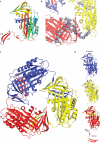
Crystal structure of the α1-antitrypsin trimer. (A) A ribbon diagram of the monomer comprising the asymmetric unit is shown coloured from N to C terminus (blue to red). The reactive centre loop is fully inserted as the fourth strand of β-sheet A (strands are numbered), and elements comprising the very C terminus, strands 1C and 4 and 5B, are indicated. (B) Clear electron density (blue wire for 2Fo−Fc, contoured at 1σ, surrounding residues 357–393, depicted as sticks) links three crystallographically related monomers into the trimer shown in (C), with each monomer in a different colour. (D) A model of an open trimer, coloured as in (C), was created by breaking one intermolecular contact and orienting the protomers in a head-to-tail manner. The red monomer is able to donate the C terminus (donor) and the blue monomer can accept a C terminus (acceptor) to elongate or self-terminate. The lightning bolt indicates a site of likely proteolytic susceptibility.
Figure 3
Figure 3
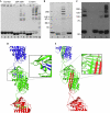
Disulphide-trapping experiments in vitro and in vivo. (A) A non-reducing SDS gel of α1-antitrypsin (α1AT) variants before and after polymerization reports the formation of intermolecular disulphide bonds as ladders. Control (Cys-free background C232S), strand 5A–6A disulphide (s5A–s6A) and C-terminal disulphide (C-term) variants are indicated. In each case, the first lane is of unincubated variant (lanes 1, 5 and 9) and the other lanes are incubations in the presence of dithiothreitol at 4°C (lanes 2, 6 and 10), 37°C with 0.75 M GndHCl (lanes 3, 7 and 11) and without GndHCl at 53°C (lanes 4, 8 and 12). After incubation, samples were dialysed and oxidized to covalently link adjacent Cys residues. Lane 13 comprises molecular mass standards (55.4, 66.3, 97.4, 116.3 and 200 kDa). (B) Non-reducing SDS gel of purified Z–α1AT polymers from P. pastoris (molecular mass standards in kDa are indicated). Lane 1 is the Cys-free background used for all variants (C232A); lane 2 is the s5A–s6A disulphide; lane 3 is the C-terminal disulphide; and lane 4 is a control with two distant Cys residues. (C) Western blot of non-reducing SDS gel of lysate from COS-7 cells transfected with Z–α1AT with and without the double Cys mutations (molecular mass standards in kDa are indicated). Lane 1 is the control with two distant Cys residues; lane 2 is untransfected control; lane 3 is the Cys-free control (C232A, background for all variants); lane 4 is s5A–s6A disulphide variant; and lane 5 is the C-terminal disulphide variant. An illustration of the mechanism-dependent capture of disulphide-linked C terminal (D) and s4A/s5A (E) polymers (each monomer in a different colour). The C-terminal domain-swap polymers are trapped by forming an intermolecular disulphide bond between a C-terminal Cys residue (position 392) and a Cys on strand 3 of sheet C (position 216). The s4A/s5A polymers are selectively trapped by the s5A–s6A disulphide (residues 292 and 339). Close-ups of the boxed regions are shown in the right panels.
Figure 4
Figure 4
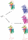
Schematic of possible folding and misfolding mechanisms of α1-antitrypsin. The unfolded state (U) is rapidly converted to an intermediate (I) that can fold either into the native conformation (N) or into a polymerigenic conformation (L*, to denote a latent-like state). L* is predictably a folding dead end in the monomeric state, but can attain the global free-energy minimum by forming polymers (P) via the C-terminal domain-swap mechanism.

Comment in

Similar articles

Cited by

References

    1. Davies MJ, Lomas DA (2008) The molecular aetiology of the serpinopathies. Int J Biochem Cell Biol 40: 1273–1286 - PubMed
    1. DeLano WL (2002) The PyMOL Molecular Graphics System (on World Wide Web) http://www.pymol.org
    1. Ekeowa UI, Freeke J, Miranda E, Gooptu B, Bush MF, Perez J, Teckman J, Robinson CV, Lomas DA (2010) Defining the mechanism of polymerization in the serpinopathies. Proc Natl Acad Sci USA 107: 17146–17151 - PMC - PubMed
    1. Emsley P, Cowtan K (2004) Coot: model-building tools for molecular graphics. Acta Crystallogr D Biol Crystallogr 60: 2126–2132 - PubMed
    1. Gooptu B, Lomas DA (2009) Conformational pathology of the serpins: themes, variations, and therapeutic strategies. Annu Rev Biochem 78: 147–176 - PubMed

Publication types

Substances

Associated data