. 2011 Sep 11;6(10):658-67.
doi: 10.1038/nnano.2011.105.

Thermodynamically stable RNA three-way junction for constructing multifunctional nanoparticles for delivery of therapeutics

Affiliations

Thermodynamically stable RNA three-way junction for constructing multifunctional nanoparticles for delivery of therapeutics

Dan Shu et al. Nat Nanotechnol. .

Abstract

RNA nanoparticles have applications in the treatment of cancers and viral infection; however, the instability of RNA nanoparticles has hindered their development for therapeutic applications. The lack of covalent linkage or crosslinking in nanoparticles causes dissociation in vivo. Here we show that the packaging RNA of bacteriophage phi29 DNA packaging motor can be assembled from 3-6 pieces of RNA oligomers without the use of metal salts. Each RNA oligomer contains a functional module that can be a receptor-binding ligand, aptamer, short interfering RNA or ribozyme. When mixed together, they self-assemble into thermodynamically stable tri-star nanoparticles with a three-way junction core. These nanoparticles are resistant to 8 M urea denaturation, are stable in serum and remain intact at extremely low concentrations. The modules remain functional in vitro and in vivo, suggesting that the three-way junction core can be used as a platform for building a variety of multifunctional nanoparticles. We studied 25 different three-way junction motifs in biological RNA and found only one other motif that shares characteristics similar to the three-way junction of phi29 pRNA.


Figures

Figure 1
Figure 1. Sequence and secondary structure of phi29 DNA-packaging RNA (pRNA)
a, Illustration of the phi29 packaging motor geared by six pRNAs (orange, pink, light blue, green, dark blue and yellow structures). b, Schematic showing a pRNA hexamer assembled through hand-in-hand interactions of six pRNA monomers. c, Sequence of pRNA monomer Ab′. Green box: central 3WJ domain. Uppercase and lowercase letters in Ab′ represent right- and left-hand loops, respectively. d, The 3WJ domain composed of three RNA oligomers in black, blue and red. Helical segments are represented as H1, H2, H3. e, A trivalent RNA nanoparticle consisting of three pRNA molecules bound at the 3WJ-pRNA core sequence (red, black and blue) and its accompanying AFM images (f). Ab′ indicates non-complementary loops.
Figure 2
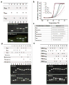
Figure 2. Assembly and stability studies of 3WJ-pRNA
In tables, ‘+’ indicates the presence of the strand in samples of the corresponding lanes. a, 15% native PAGE showing the assembly of the 3WJ core, stained by EB (upper) and SYBR Green II (lower). b, Melting curves for the assembly of the 3WJ core. The melting curves for the individual strands (brown, green, silver), two-strand combinations (blue, cyan, pink), and three-strand combination (red) are shown. c, Oligo sequences of 3WJ-pRNA cores and mutants. “del U”-deletion of U bulge; “del UUU”-deletion of UUU bulge; “del 4-nt”-deletion of 2-nucleotides at 3′ and 5′ ends, respectively. d, Length requirements for the assembly of 3WJ cores and stability assays by urea denaturation. e, Comparison of DNA and RNA 3WJ-core in native and urea gel.
Figure 3
Figure 3. Competition and dissociation assays of 3WJ-pRNA
a, Temperature effects on the stability of 3WJ-pRNA core, denoted [ab*c]3WJ evaluated by 16% native gel. Fixed concentration of Cy3 labeled [ab*c]3WJ was incubated with varying concentration of unlabeled b3WJ at 25°C, 37°C, and 55°C. b, Urea denaturing effects on the stability of [ab*c]3WJ evaluated by 16% native gel. Fixed concentration of labeled [ab*c]3WJ was incubated with unlabelled b3WJ at ratios of 1:1 and 1:5 in the presence of 0–6M urea at 25°C. c, Dissociation assay for the [32P]-3WJ-pRNA complex harboring three monomeric pRNA by 2-fold serial dilution (lanes 1–9). Monomer unit is shown on the left.
Figure 4
Figure 4. Construction of multi-module RNA nanoparticles harboring siRNA, ribozyme, and aptamer
a-c, Assembly of RNA nanoparticles with functionalities using the 3WJ-pRNA and 3WJ-5S rRNA as scaffolds. a, Illustration; b, 8% native (upper) and denaturing (lower) PAGE gel; c, AFM images of 3WJ-pRNA-siSur-rZ-FA nanoparticles. d–e, Assessing the catalytic activity of the Hepatitis B virus (HBV) ribozyme incorporated into the 3WJ-pRNA (d) and 3WJ-5S rRNA (e) cores, evaluated in 10% 8M Urea PAGE. The cleaved RNA product is boxed. Positive control: pRNA/HBV-Rz; Negative control: 3WJ-RNA/siSur-MG-FA. f–g, Functional assay of the MG (malachite green) aptamer incorporated in RNA nanoparticles using the 3WJ-pRNA (f) and 3WJ-5S rRNA (g) cores. MG fluorescence was measured using excitation wavelengths 475 and 615nm.
Figure 5
Figure 5. In vitro and in vivo binding and entry of 3WJ-pRNA nanoparticles into targeted cells
a, Flow cytometry revealed the binding and specific entry of fluorescent-[3WJ-pRNA-siSur-rZ-FA] nanoparticles into folate receptor positive (FA+) cells. Positive and negative controls were Cy3-FA-DNA and Cy3-[3WJ-pRNA-siSur-rZ-NH2] (without FA), respectively. b, Confocal images showed targeting of FA+-KB cells by co-localization (overlap, 4) of cytoplasma (green, 1) and RNA nanoparticles (red, 2) (magnified, bottom panel). Blue–nuclei, 3. c–d, Target gene knock-down effects showed by (c) qRT-PCR with GADPH as endogenous control and by (d) Western blot assay with β–actin as endogenous control. e, 3WJ-pRNA nanoparticles target FA+ tumor xenografts upon systemic administration in nude mice. Upper panel: whole body; Lower panel: organ imaging; Lv=liver; K=kidney; H=heart; L=lung; S=spleen; I=intestine; M=muscle; T=tumor). Scale: Fluorescent Intensity.
Figure 6
Figure 6. Comparison of different 3WJ-RNA core
a, Assembly and stability of 11 3WJ-RNA core motifs assayed in 16% native (upper) and 16% 8M Urea (lower) PAGE gel. b, Melting curves for each of the 11 RNA 3WJ core motifs assembled from three oligos for each 3WJ motif under the physiological buffer TMS. Please refer to the Table 1 for the respective Tm values.

Similar articles

Cited by

References

    1. Seeman NC. Nanomaterials based on DNA. Annu Rev Biochem. 2010;79:65–87. - PMC - PubMed
    1. Guo P. The emerging field of RNA nanotechnology. Nat Nanotechnol. 2010;5:833–842. - PMC - PubMed
    1. Guo P, et al. Inter-RNA interaction of phage phi29 pRNA to form a hexameric complex for viral DNA transportation. Mol Cell. 1998;2:149–155. - PubMed
    1. Shukla GC, et al. A Boost for the Emerging Field of RNA Nanotechnology. ACS Nano. 2011;5:3405–3418. - PMC - PubMed
    1. Cruz JA, Westhof E. The Dynamic Landscapes of RNA Architecture. Cell. 2009;136:604–609. - PubMed

Publication types