. 2011 Sep 11;14(10):1253-9.
doi: 10.1038/nn.2908.

Ligand-binding domain of an α7-nicotinic receptor chimera and its complex with agonist

Affiliations

Ligand-binding domain of an α7-nicotinic receptor chimera and its complex with agonist

Shu-Xing Li et al. Nat Neurosci. .

Abstract

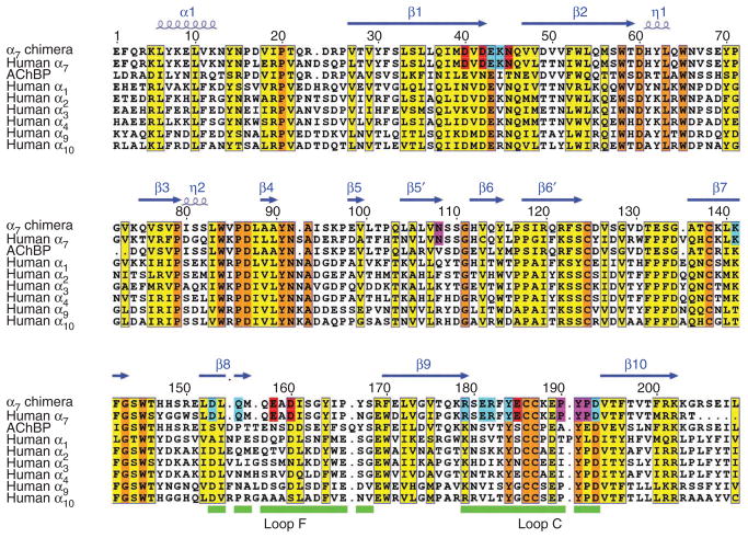
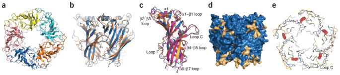
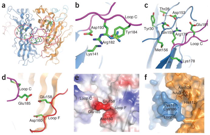
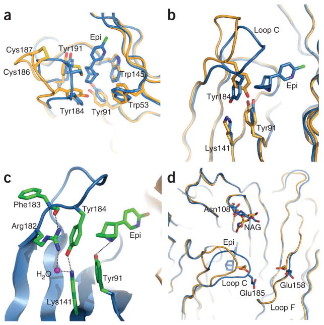
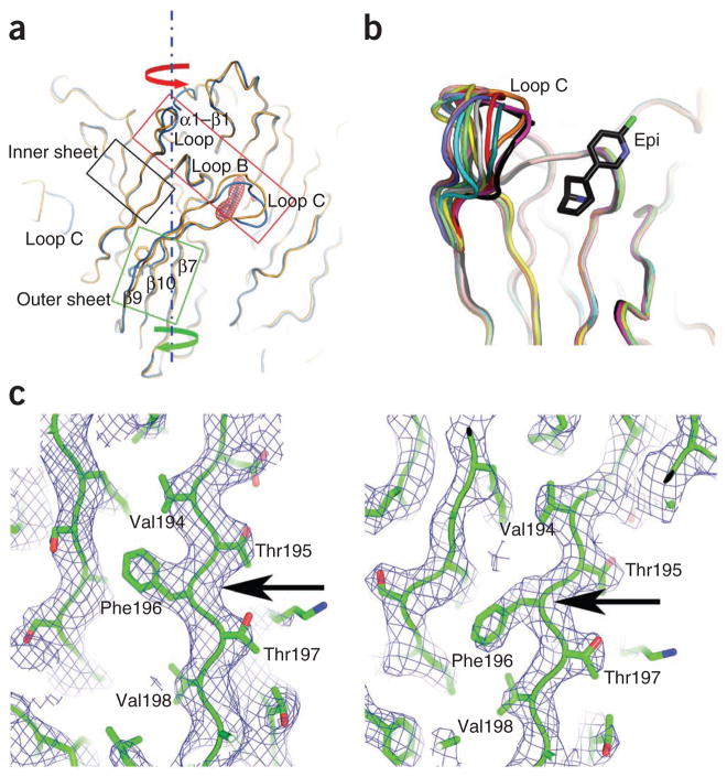
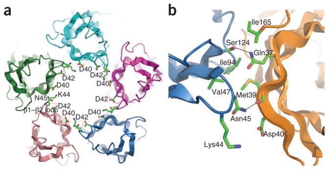
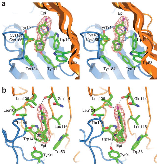
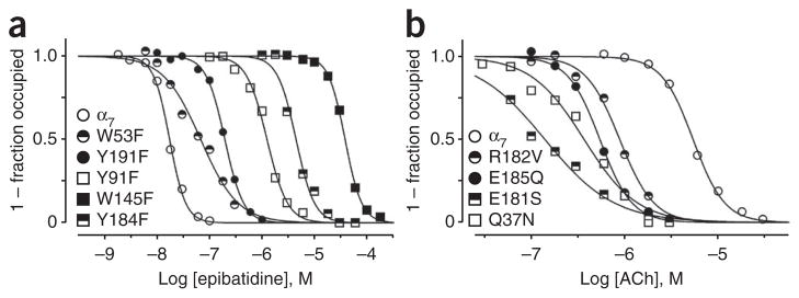
The α(7) acetylcholine receptor (AChR) mediates pre- and postsynaptic neurotransmission in the central nervous system and is a potential therapeutic target in neurodegenerative, neuropsychiatric and inflammatory disorders. We determined the crystal structure of the extracellular domain of a receptor chimera constructed from the human α(7) AChR and Lymnaea stagnalis acetylcholine binding protein (AChBP), which shares 64% sequence identity and 71% similarity with native α(7). We also determined the structure with bound epibatidine, a potent AChR agonist. Comparison of the structures revealed molecular rearrangements and interactions that mediate agonist recognition and early steps in signal transduction in α(7) AChRs. The structures further revealed a ring of negative charge within the central vestibule, poised to contribute to cation selectivity. Structure-guided mutational studies disclosed distinctive contributions to agonist recognition and signal transduction in α(7) AChRs. The structures provide a realistic template for structure-aided drug design and for defining structure-function relationships of α(7) AChRs.


Conflict of interest statement

COMPETING FINANCIAL INTERESTS

The authors declare no competing financial interests.

Figures

Figure 1
Figure 1
Sequence and numbering of the α7–AChBP chimera and its alignment with related AChR sequences. Orange indicates invariant residues and yellow indicates partially conserved residues. Secondary structures are shown schematically above the sequences. Putative functionally important residues for ligand recognition (pink), signal transduction (blue) and inorganic ion binding (red) are shown. Loops F and C are indicated by green bars.
Figure 2
Figure 2
Overall structures of the α7–AChBP chimera and comparison to related structures. (a) Top view of the α7–AChBP chimera pentamer along the five-fold axis of symmetry; each subunit is shown in a different color. (b) Structure superposition between the α7–AChBP chimera (blue) and AChBP (orange) pentamers viewed from the side that is normal to the five-fold axis. (c) Structure superposition of subunits from the α7–AChBP chimera (blue), α1 extracellular domain (magenta) and AChBP (orange); loops showing substantial differences are labeled. (d) Surface representation showing α7 residues (blue) and AChBP residues area (beige) on the α7–AChBP chimera. (e) Backbone superposition between the Apo (gold) and Epi (blue) structures viewed down the five-fold axis. The epibatidine molecule (Epi) is shown by the FoFc electron density contoured at the 3.0-σ level.
Figure 3
Figure 3
Structures specific to α7 revealed by the α7–AChBP chimera. (a) Four regions of α7-specific residues near loops C (magenta) and F (red), indicated by I–IV. (b) Close-up of the signal transduction region beneath loop C. Alternative conformations of Arg182 are indicated by different colors. (c) Close-up of linkage region between loops C and F within the same subunit. (d,e) Glu185-Glu158-Asp160 triad spanning loop C of the principal subunit and loop F of the complementary subunit, in ribbon (d) and surface (e) representation. Positive and negative surface potentials are indicated by blue and red, respectively. (f) Close-up of glycan across from loop C. NAG, N-acetylglucosamine.
Figure 4
Figure 4
Epibatidine-induced conformational changes. (a) Backbone superposition between the Apo (gold) and Epi (blue) structures shows a clockwise rotation of the outer β-sheet (green box and arrow, bottom) and a counterclockwise rotation of the top part of the subunit structure (red box and arrow, top) when viewed down the pentamer axis. The stationary inner sheet is indicated by the black box, and the epibatidine molecule is shown in electron density. The side chain rotamer switch of Phe196 is also evident in the green box. (b) Backbone superposition of individual subunits show variable conformations of loop C in the Apo structure (ten subunits colored differently) but a single closed conformation in the Epi structure (black, only one structure shown). Epi indicates the epibatidine molecule. (c) The 2FoFc electron density map (contoured at the 1.0-σ level) shows the distinct side chain conformations of Phe196 (arrows) in the Apo (left) and Epi (right) structures, demonstrating repacking of the protein core as a result of epibatidine-induced structural changes.
Figure 5
Figure 5
Epibatidine-induced structural reorganization of the ligand-binding pocket and flanking regions. (a) Comparison of the ligand-binding pocket between the Apo (gold) and Epi (blue) structures. (b) Comparison of key residues underneath loop C implicated in signal transduction. (c) Highly ordered assembly of Tyr184, Tyr91, Lys141, Arg182 and a solvent molecule in the Epi structure. Epi indicates the epibatidine molecule. (d) Comparison of the interactions at the tip of loop C between the Apo (gold) and Epi (blue) structures. NAG, N-acetylglucosamine.
Figure 6
Figure 6
Molecular recognition of epibatidine. (a) Stereo view of the ligand-binding pocket from the side of the pentamer showing the position of epibatidine (Epi) in the aromatic cage. The protein is in ribbon style and the epibatidine molecule is shown with the FoFc electron density contoured at the 3.0-σ level. (b) Stereo view of the ligand-binding pocket from above the pentamer. This view highlights hydrogen-bond interactions and interactions with the complementary face of the binding site between epibatidine and the receptor chimera.
Figure 7
Figure 7
Pore-facing regions of inter-subunit contact. (a) Bottom view of the pentamer along the five-fold axis of symmetry, showing the tandem arrangement of the β1–β2 loops and the ring of ten aspartate and five asparagine residues. (b) Inter-subunit contacts between the tip of the β1–β2 loop of the principal subunit (blue) and the stem of the β1–β2 loop of the complementary subunit (orange).
Figure 8
Figure 8
Agonist binding after mutation of key residues. (a) Ligand contact residues. (b) Non-contact residues. Binding of epibatidine and ACh to native α7 AChRs was measured by competition against the initial rate of 125I-labeled α-bungarotoxin binding (see Online Methods). Curves are nonlinear least-squares fits of the Hill equation to the data with fit parameters given in Supplementary Table 2.

Similar articles

Cited by

References

    1. Brejc K, et al. Crystal structure of an ACh-binding protein reveals the ligand-binding domain of nicotinic receptors. Nature. 2001;411:269–276. - PubMed
    1. Celie PH, et al. Nicotine and carbamylcholine binding to nicotinic acetylcholine receptors as studied in AChBP crystal structures. Neuron. 2004;41:907–914. - PubMed
    1. Hansen SB, et al. Structures of Aplysia AChBP complexes with nicotinic agonists and antagonists reveal distinctive binding interfaces and conformations. EMBO J. 2005;24:3635–3646. - PMC - PubMed
    1. Unwin N. Refined structure of the nicotinic receptor at 4 Å resolution. J Mol Biol. 2005;346:967–989. - PubMed
    1. Dellisanti CD, Yao Y, Stroud JC, Wang ZZ, Chen L. Crystal structure of the extracellular domain of nAChR α1 bound to alpha-bungarotoxin at 1.94 Å resolution. Nat Neurosci. 2007;10:953–962. - PubMed

Publication types

MeSH terms