. 2012 Mar;9(2):147-54.
doi: 10.1038/cmi.2011.28. Epub 2011 Sep 12.

CDR3δ -grafted γ9δ2T cells mediate effective antitumor reactivity

Affiliations

CDR3δ -grafted γ9δ2T cells mediate effective antitumor reactivity

Hui Zhao et al. Cell Mol Immunol. 2012 Mar.

Abstract

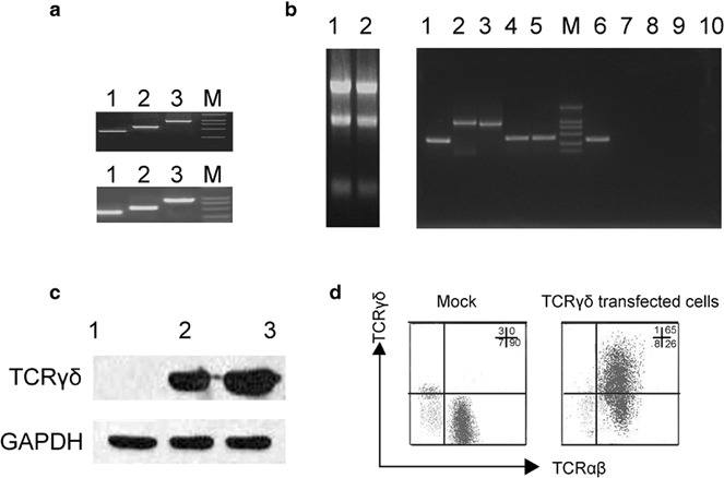
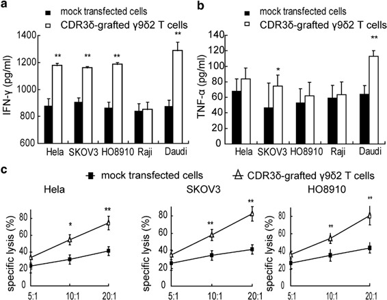
Adoptive cell-transfer therapy (ACT) has been reported to suppress growing tumors and to overcome tumor escape in animal models. As a candidate ACT effector, γ9δ2T cells can be activated and expanded in vitro and in vivo and display strong antitumor activity against colorectal, lung, prostate, ovarian and renal cell carcinomas. However, it is difficult to obtain a large enough number of γδT cells to meet the need for immunotherapy that can overcome the cancer patients' immune suppressive tumor microenvironment. In previous studies, our lab confirmed that γ9δ2T cells recognized tumor cells via the CDR3δ region of the γδ-T-cell receptor (TCR). We constructed full-length human peripheral blood mononuclear cell (PBMC)-derived γ9 and δ2 chains in which the CDR3 region was replaced by an ovarian epithelial carcinoma (OEC)-derived CDR3. We transferred the CDR3δ-grafted γ9δ2TCR into peripheral blood lymphocytes (PBLs) to develop genetically modified γ9δ2T cells. In vitro studies have shown that these CDR3δ-grafted γ9δ2T cells can produce cytokines after stimulation with tumor cell extracts and exhibit cytotoxicity towards tumor cells, including human OEC and cervical adenocarcinoma. CDR3δ-grafted γ9δ2T cells adoptively transferred into nude mice bearing a human OEC cell line demonstrated significant antitumor effects. These results indicate that CDR3δ-grafted γ9δ2T cells might be candidates for clinical tumor immunotherapy.


Figures

Figure 1
Figure 1
Transfected PBLs expressed the γδTCR with a tumor antigen-specific CDR3δ sequence. (a) PCR products of the full-length γ9 and δ2 chains containing the tumor-specific CDR3 sequence. The upper image displays the PCR products of the full-length γ9 chain. Lane 1: the first half sequence of the full-length γ9 chain; lane 2: the second half of the full-length γ9 chain; lane 3: the full-length γ9 chain. The lower image shows the PCR products of the full-length δ2 chain containing the tumor antigen-specific CDR3 sequence derived from TILs isolated from OEC. Lane 1: the first half sequence of the full length δ2 chain containing the OEC-derived CDR3 sequence; lane 2: the second half of the full length δ2 chain with the OEC-derived CDR3 sequence; lane 3: the full length δ2 chain with the OEC-derived CDR3 sequence. (b) PBLs isolated from a healthy donor were stimulated for 3 days and then transfected with a retrovirus containing pMSCVhyg-γ9 and pMSCVneo-δ2 (OT3) in culture media containing 200 IU/ml IL-2. The expression of the full-length γ9 and δ2 chains and their respective CDR3 region mRNA in transfected cells was analyzed by RT-PCR. Left: the mRNA extracted from mock-transfected cells (lane1) and TCR-transfected cells (lane2); right: the β-actin from TCR-transfected cells (lane1);, the full-length γ9 chain from the TCR-transfected cells (lane 2); the full-length δ2 chain from the TCR-transfected cells (lane 3); the CDR3 region of the γ9 chain from the TCR-transfected cells (lane 4); the CDR3 region of the δ2 chain from the TCR-transfected cells (lane 5); β-actin expression in the mock-transfected cells (lane 6); the full-length γ9 chain from the mock-transfected cells (lane 7); the full-length δ2 chain from the mock-transfected cells (lane 8); the CDR3 region of the γ9 chain from the mock-transfected cells (lane 9); and the CDR3 region of the δ2 chain from the mock-transfected cells (lane 10). (c) Western blot analysis of γδTCR expression. Lane 1: mock control; lane2: TCR-transfected cells; lane 3: γδT cells as a positive control. (d) Surface expression of the γδTCR on transfected cells was analyzed by FCM. The cells were stained with FITC-conjugated anti-γδTCR mAbs and polyethylene-conjugated anti-αβTCR mAbs. γδTCR and αβTCR cell surface expression on mock-transfected cells (left) and TCRγδ-transfected cells (right) was determined by flow cytometry. One representative result from three independent experiments is shown. FCM, flow cytometry; GAPDH, glyceraldehyde 3-phosphate dehydrogenase; OEC, ovarian epithelial carcinoma; PBL, peripheral blood lymphocyte; RT, reverse transcription; TCR, T-cell receptor; TIL, tumor-infiltrating lymphocyte.
Figure 2
Figure 2
Tumor cell recognition and cytotoxicity by CDR3δ-grafted γ9δ2T cells. (a) The CDR3δ-grafted γ9δ2T cells or mock-transfected cells were incubated with tumor cell extracts from HeLa, SKOV3, HO8910, Daudi and Raji cell lines for 72 h, after which the supernatant was harvested and IFN-γ production was measured by ELISA. (b) The CDR3δ-grafted γ9δ2T cells or mock-transfected cells were incubated with HeLa, SKOV3, HO8910, Daudi and Raji cell extracts for 24 h, after which the supernatant was harvested for detection of TNF-α production by ELISA. (c) The MTT assay was used to evaluate the transfected cell cytotoxicity against tumor cells. The transfected effector cells were incubated with HeLa, SKOV3 and HO8910 target cells at different effector-to-target ratios for 8 h MTT solution (5 mg/ml) was added to each well (10 µl/well) and cells were incubated at 37 °C for an additional 4 h. The reaction was stopped by the addition of 100 µl DMSO to dissolve the tetrazolium crystals. The plate was examined at 570nm with 630nm correction  in a Multiskan Microplate Reader. Asterisks indicate significant differences compared to the mock control: *P<0.05, **P<0.01. All data are representative of three independent experiments. DMSO, dimethylsulfoxide; IFN, interferon; MTT, 3-(4,5-dimethyl-thiazol-2-yl)-2,5-diphenyltetrazolium bromide.
Figure 3
Figure 3
The antitumor effects of CDR3δ-grafted γ9δ2T cells were blocked by an anti-TCRγδ mAb. (a) CDR3δ-grafted γδT cells were incubated with an anti-TCRγδ mAb, a mouse IgG1 isotype control or no mAb for 2 h and then stimulated with tumor cell extracts for 72 h. IFN-γ secretion in the supernatants was detected by ELISA. (bd)The CDR3δ-grafted γ9δ2T cells were incubated with an anti-TCRγδ mAb, mouse IgG1 isotype control or no treatment for 2 h and then cocultured with tumor target cells at different E/T ratios. The MTT assay was used to evaluate cytotoxicity. Asterisks indicated significant differences compared to the mock control, *P<0.05, **P<0.01. The results are representative of three independent experiments. E/T, effector/target; IFN, interferon; mAb, monoclonal antibody; MTT, 3-(4,5-dimethylHSthiazol-2-yl)-2,5-diphenyltetrazolium bromide; TCR, T-cell receptor.
Figure 4
Figure 4
The CDR3δ-grafted γ9δ2T antitumor cytotoxicity mechanism. (a) Influence of treatment with an anti-Fas mAb on cytotoxicity of CDR3δ-grafted γ9δ2T cells against various tumor cell lines. Tumor cells were treated with an anti-Fas mAb at saturating concentrations for 2 h before being incubated with effector cells. (b) Effects of BFA on CDR3δ-grafted γ9δ2T-cell antitumor cytotoxicity. CDR3δ-grafted γ9δ2T cells were treated with BFA at saturating concentrations for 2 h before being incubated with target tumor cells (HeLa, SKOV3 and HO8910). (c) Effects of CMA on CDR3-grafted γ9δ2T-cell antitumor cytotoxicity. CDR3δ-grafted γ9δ2T cells were incubated with CMA at saturating concentrations for 2 h before the cytotoxicity assay. (d) CDR3δ-grafted γ9δ2T cells were pretreated with a combination of CMA (100 nM) and BFA (25 µM) before being cocultured with tumor cells at an E/T ratio of 10:1. **P<0.01, no treatment group vs. CMA-treated group; # # P<0.01, no treatment group vs. CMA+BFA-treated group. The results are representative of three independent experiments. BFA, brefeldin A; CMA, concanamycin A; E/T, effector/target; mAb, monoclonal antibody.
Figure 5
Figure 5
In vivo antitumor effects of CDR3-grafted γ9δ2T cells. Mice were injected subcutaneously 1×10⁶ SKOV3 cells in the back right flank. When the tumor mass grew to approximately 100 mm3, CDR3δ-grafted γ9δ2T cells, mock-transfected cells or PBS were injected intratumorally every 3 days for a total of four injections. Human IL-2 was administered intraperitoneally at a dose of 5000 IU for all mice with the exception of the PBS group. The general condition of nude mice and tumor progression were observed every 2 days. Mouse survival was observed every day. (a) Mice treated with CDR3δ-grafted γ9δ2T cells showed slow tumor mass growth compared to mice treated with mock-transfected cells or PBS (P<0.05), whereas mock-transfected cells-treated mice and PBS-treated mice displayed similar tumor progression (P>0.05). (b) Survival of nude mice bearing SKOV3 cells was prolonged by treatment with CDR3-grafted γ9δ2T cells plus IL-2. Treatment with CDR3-grafted γ9δ2T cells resulted in longer survival when compared to PBS treated group or mock-transfected group (P<0.05). PBS, phosphate-buffered saline.

Similar articles

Cited by

References

    1. Peng L, Shu SKrauss JC. Treatment of subcutaneous tumor with adoptively transferred T cells. Cell Immunol. 1997;178:24–32. - PubMed
    1. Rosenberg SA, Spiess P, Lafreniere R. A new approach to the adoptive immunotherapy of cancer with tumor-infiltrating lymphocytes. Science. 1986;233:1318–1321. - PubMed
    1. Plautz GE, Bukowski RM, Novick AC, Klein EA, Kursh ED, Olencki TE, et al. T-cell adoptive immunotherapy of metastatic renal cell carcinoma Urology 199954617–623.discussion 623–614. - PubMed
    1. Dudley ME, Wunderlich J, Nishimura MI, Yu D, Yang JC, Topalian SL, et al. Adoptive transfer of cloned melanoma-reactive T lymphocytes for the treatment of patients with metastatic melanoma. J Immunother. 2001;24:363–373. - PubMed
    1. Dudley ME, Rosenberg SA. Adoptive-cell-transfer therapy for the treatment of patients with cancer. Nat Rev Cancer. 2003;3:666–675. - PMC - PubMed

Publication types

MeSH terms

Substances