. 2012 Apr 26;31(17):2237-46.
doi: 10.1038/onc.2011.392. Epub 2011 Sep 12.

Loss of breast epithelial marker hCLCA2 promotes epithelial-to-mesenchymal transition and indicates higher risk of metastasis

Affiliations

Loss of breast epithelial marker hCLCA2 promotes epithelial-to-mesenchymal transition and indicates higher risk of metastasis

V Walia et al. Oncogene. .

Abstract

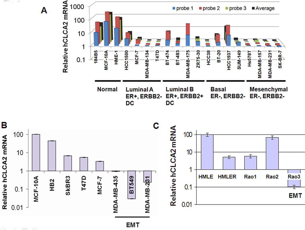
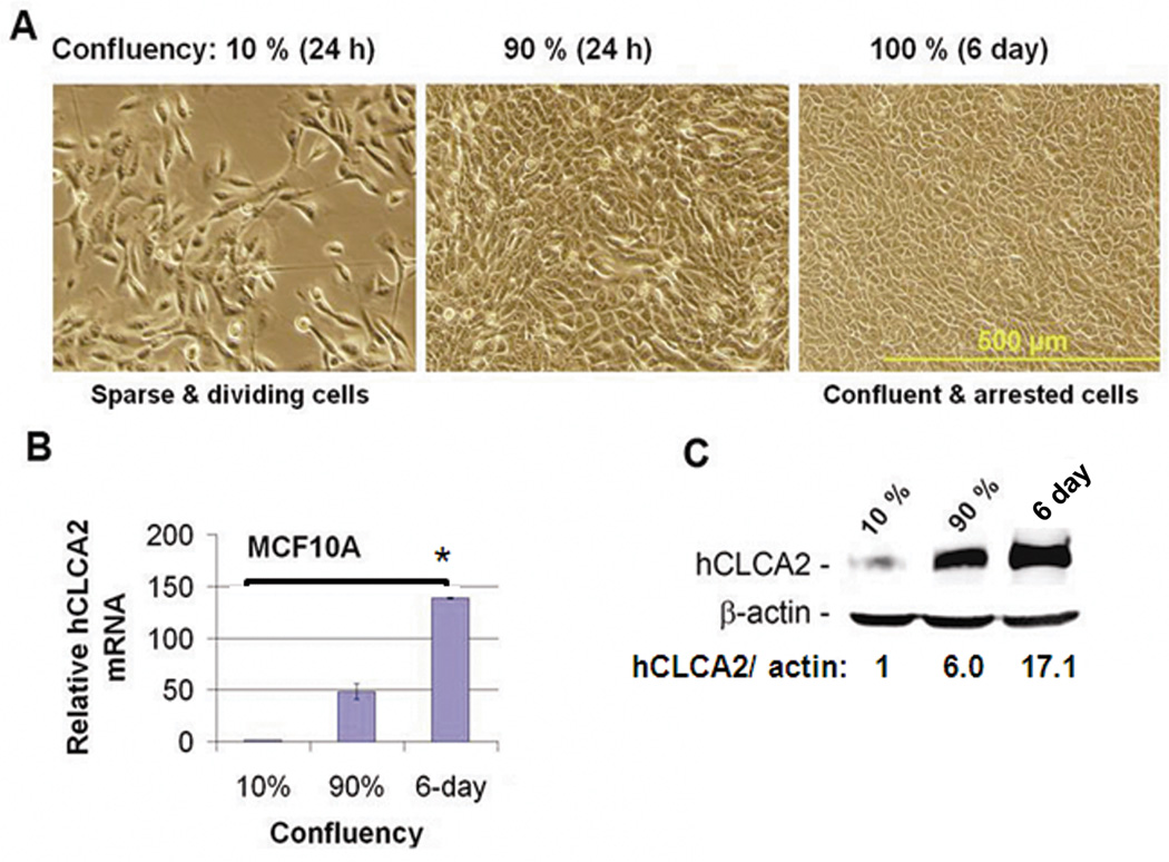
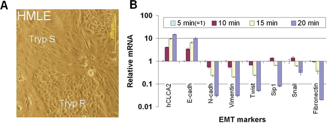
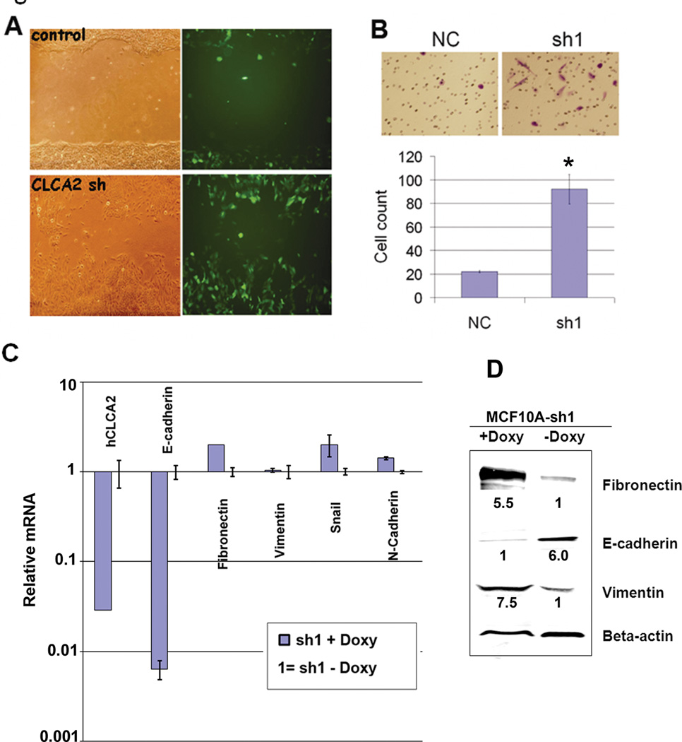
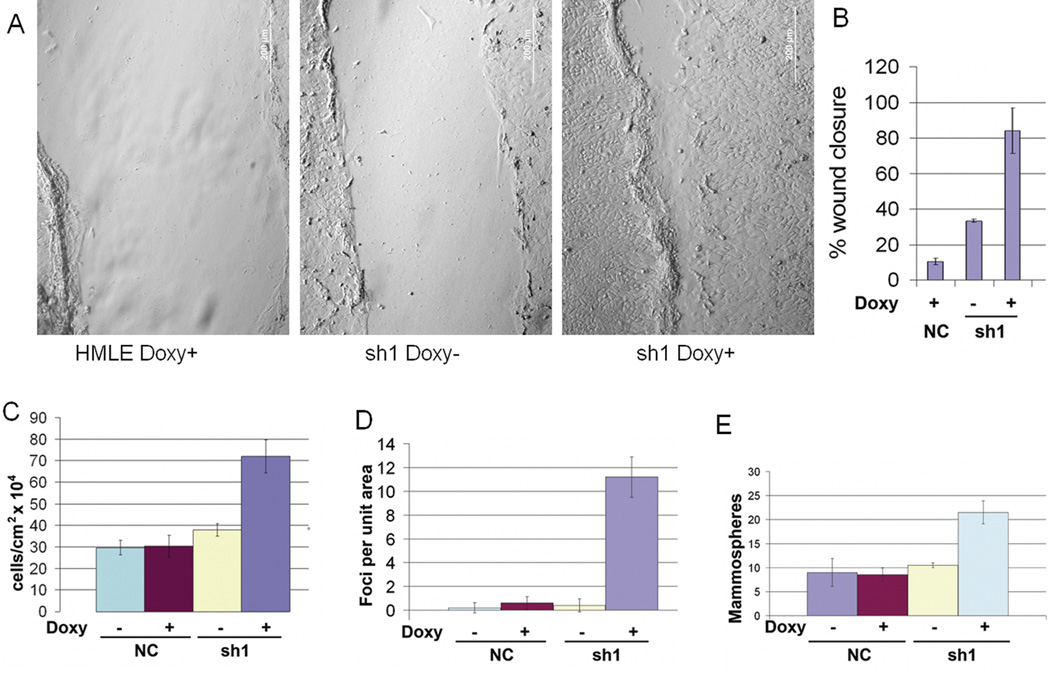
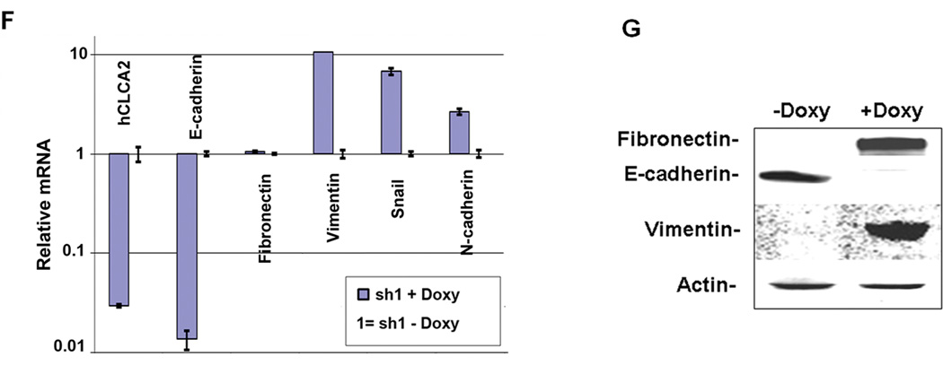
Transition between epithelial and mesenchymal states is a feature of both normal development and tumor progression. We report that expression of chloride channel accessory protein hCLCA2 is a characteristic of epithelial differentiation in the immortalized MCF10A and HMLE models, while induction of epithelial-to-mesenchymal transition by cell dilution, TGFβ or mesenchymal transcription factors sharply reduces hCLCA2 levels. Attenuation of hCLCA2 expression by lentiviral small hairpin RNA caused cell overgrowth and focus formation, enhanced migration and invasion, and increased mammosphere formation in methylcellulose. These changes were accompanied by downregulation of E-cadherin and upregulation of mesenchymal markers such as vimentin and fibronectin. Moreover, hCLCA2 expression is greatly downregulated in breast cancer cells with a mesenchymal or claudin-low profile. These observations suggest that loss of hCLCA2 may promote metastasis. We find that higher-than-median expression of hCLCA2 is associated with a one-third lower rate of metastasis over an 18-year period among breast cancer patients compared with lower-than-median (n=344, unfiltered for subtype). Thus, hCLCA2 is required for epithelial differentiation, and its loss during tumor progression contributes to metastasis. Overexpression of hCLCA2 has been reported to inhibit cell proliferation and is accompanied by increases in chloride current at the plasma membrane and reduced intracellular pH (pHi). We found that knockdown cells have sharply reduced chloride current and higher pHi, both characteristics of tumor cells. These results suggest a mechanism for the effects on differentiation. Loss of hCLCA2 may allow escape from pHi homeostatic mechanisms, permitting the higher intracellular and lower extracellular pH that are characteristic of aggressive tumor cells.


Conflict of interest statement

Conflict of Interest

The authors declare no conflict of interest.

Figures

Figure 1
Figure 1
Downregulation of hCLCA2 in breast cancer cell lines. A, transcriptional profile data extracted from an Affymetrix cDNA microarray study of breast cancer cell lines (Charafe-Jauffret, Ginestier et al. 2006). B, RT-qPCR data confirming that mesenchymal cell lines have lowest expression. C, expression profile of cancer cell lines derived by genetic modification in vitro.
Figure 2
Figure 2
Expression of hCLCA2 is induced at high cell density. Cells were seeded at 10% or 90% confluency and incubated for 24h or 6 days past confluency before measuring expression levels of hCLCA2 mRNA or protein relative to internal standards. A, phase-contrast images. Scale bar indicates 500 µm. B, quantification of hCLCA2 mRNA by RT-qPCR. P <0.05. C, quantification of protein induction by immunoblot. The fold increase in hCLCA2 is indicated after normalization to actin internal control.
Figure 3
Figure 3
hCLCA2 expression correlates with epithelial differentiation. HMLE cells were separated into epithelioid and mesenchymal subpopulations by differential trypsinization and subjected to RT-qPCR. A, micrograph showing typical cobblestone-like epithelial islands surrounded by elongated mesenchymal cells. B, transcriptional profile by RT-qPCR showing that hCLCA2 expression peaks in the most trypsin-resistant fraction, correlating directly with E-cadherin but inversely with mesenchymal markers. Expression in the 5 min pool was normalized as 1.
Figure 4
Figure 4
Repression of hCLCA2 by mesenchymal transcription factors and TGF beta. A, immunoblot showing repression of both hCLCA2 and E-cadherin in MCF10A infected with an adenovirus encoding Snail. Relative expression levels are indicated after normalization to actin. B, repression of hCLCA2 in HMLE transduced with lentiviruses encoding Snail, Twist, Goosecoid, or TGF beta. C, repression of hCLCA2 in MCF10A treated with 2.5 ng/ml TGF beta relative to untreated control (=1). B and C show RT-qPCR data.
Figure 5
Figure 5
Knockdown of hCLCA2 imparts a migratory and invasive phenotype to MCF10A. A, wound assays conducted with MCF10A cells transduced with GIPZ bearing hCLCA2 shRNA or control insert. B, Matrigel invasion assay in a Boyden chamber. N = 3, p <= 0.0006489. C, transcriptional profile of EMT markers in cells expressing shRNA1 in a doxycycline-dependent manner from TRIPZ vector. Doxycycline had no effect on vector-transduced controls. D, expression of EMT marker proteins in cell lysates. Relative expression levels are indicated normalized to actin.
Figure 6
Figure 6
Knockdown of hCLCA2 in HMLE promotes the loss of epithelial differentiation. HMLE cells were transduced with TRIPZ lentivirus expressing hCLCA2 shRNA1 in a doxycycline-dependent manner. Control cells contained a non-silencing control treated with doxycycline (first panel) or untreated. A, wound assays in presence or absence of doxycycline. B, quantification of wound closure. C, cell number 4 days after seeding at 100% confluency. D, focus formation. E, increased mammosphere formation by hCLCA2 knockdown cells. F, transcriptional profile of EMT markers. G, immunoblots of whole-cell lysates. P values for sh1, minus vs. plus doxycycline, for B, C, D, and E respectively were 0.000431, 0.006891, 0.0002145, and 0.001748; n = 3.
Figure 6
Figure 6
Knockdown of hCLCA2 in HMLE promotes the loss of epithelial differentiation. HMLE cells were transduced with TRIPZ lentivirus expressing hCLCA2 shRNA1 in a doxycycline-dependent manner. Control cells contained a non-silencing control treated with doxycycline (first panel) or untreated. A, wound assays in presence or absence of doxycycline. B, quantification of wound closure. C, cell number 4 days after seeding at 100% confluency. D, focus formation. E, increased mammosphere formation by hCLCA2 knockdown cells. F, transcriptional profile of EMT markers. G, immunoblots of whole-cell lysates. P values for sh1, minus vs. plus doxycycline, for B, C, D, and E respectively were 0.000431, 0.006891, 0.0002145, and 0.001748; n = 3.
Figure 7
Figure 7
Effect of hCLCA2 expression level on metastasis-free survival in breast cancer patients. Transcriptional profiles of primary breast tumors were analyzed for hCLCA2 expression and related to metastasis incidence over an 18 year period. Data are presented as a Kaplan-Meier plot of metastasis-free survival (proportion vs. time) of 344 patients with high (blue line) or low (red line) expression level of the gene "CLCA2". Chi-square p-value = 0.00428.

Similar articles

Cited by

References

    1. Agiostratidou G, Li M, et al. Loss of retinal cadherin facilitates mammary tumor progression and metastasis. Cancer Res. 2009;69(12):5030–5038. - PMC - PubMed
    1. Antonsson B, Conti F, et al. Inhibition of Bax channel-forming activity by Bcl-2. Science. 1997;277(5324):370–372. - PubMed
    1. Barriere H, Poujeol C, et al. CFTR modulates programmed cell death by decreasing intracellular pH in Chinese hamster lung fibroblasts. Am. J. Physiol Cell Physiol. 2001;281(3):C810–C824. - PubMed
    1. Barry MA, Eastman A. Endonuclease activation during apoptosis: the role of cytosolic Ca2+ and pH. Biochem. Biophys. Res. Commun. 1992;186(2):782–789. - PubMed
    1. Beckley JR, Pauli BU, et al. Re-expression of detachment-inducible chloride channel mCLCA5 suppresses growth of metastatic breast cancer cells. J. Biol. Chem. 2004;279(40):41634–41641. - PubMed

Publication types