

Framing postpartum hemorrhage as a consequence of human placental biology: an evolutionary and comparative perspective
- PMID: 21909154
- PMCID: PMC3168987
- DOI: 10.1111/j.1548-1433.2011.01351.x
Framing postpartum hemorrhage as a consequence of human placental biology: an evolutionary and comparative perspective
Abstract
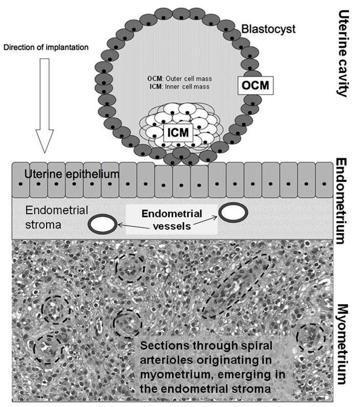
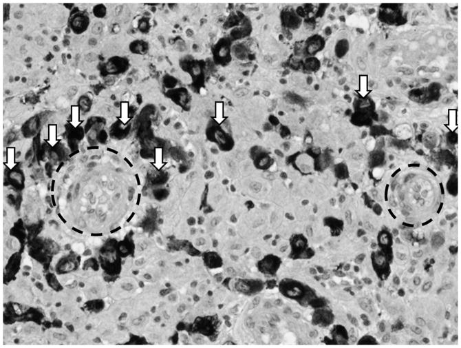
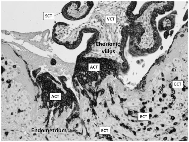
Postpartum hemorrhage (PPH), the leading cause of maternal mortality worldwide, is responsible for 35 percent of maternal deaths. Proximately, PPH results from the failure of the placenta to separate from the uterine wall properly, most often because of impairment of uterine muscle contraction. Despite its prevalence and its well-described clinical manifestations, the ultimate causes of PPH are not known and have not been investigated through an evolutionary lens. We argue that vulnerability to PPH stems from the intensely invasive nature of human placentation. The human placenta causes uterine vessels to undergo transformation to provide the developing fetus with a high plane of maternal resources; the degree of this transformation in humans is extensive. We argue that the particularly invasive nature of the human placenta increases the possibility of increased blood loss at parturition. We review evidence suggesting PPH and other placental disorders represent an evolutionarily novel condition in hominins.
Keywords: comparative placentation; evolutionary medicine; implantation; postpartum hemorrhage; trophoblast.
Figures
References
REFERENCES CITED
-
- Abitbol M Maurice. Growth of the Fetus in the Abdominal Cavity. American Journal of Physical Anthropology. 1993;91(3):367–378. - PubMed
-
- Angstmann Tobias, Gard Gregory, Harrington Tim, Ward Elizabeth, Thomson Amanda, Giles Warwick. Surgical Management of Placenta Accreta: A Cohort Series and Suggested Approach. American Journal of Obstetrics and Gynecology. 2009;202(1):38e1–38e9. - PubMed
-
- Athanassiades Andrew, Lala Peeyush K. Role of Placenta Growth Factor (PIGF) in Human Extravillous Trophoblast Proliferation, Migration and Invasiveness. Placenta. 1998;19(7):465–473. - PubMed
-
- Barker David J. In Utero Programming of Chronic Disease. Clinical Science. 1998;95(2):115–128. - PubMed
FOR FURTHER READING
-
- Fleuriet K Jill. La Tecnología y Las Monjitas: Constellations of Authoritative Knowledge at a Religious Birthing Center in South Texas. Medical Anthropology Quarterly. 2009;23(3):212–234. - PubMed
-
- Gettler Lee T. Direct Male Care and Hominin Evolution: Why Male–Child Interaction Is More Than a Nice Social Idea. American Anthropologist. 2010;112(1):7–21.
-
- Hagen Edward H, Clark Barrett H. Perinatal Sadness among Shuar Women: Support for an Evolutionary Theory of Psychic Pain. Medical Anthropology Quarterly. 2007;21(1):22–40. - PubMed
-
- Macdonald Margaret. Gender Expectations: Natural Bodies and Natural Births in the New Midwifery in Canada. Medical Anthropology Quarterly. 2006;20(2):235–256. - PubMed
-
- Wendland Claire L. The Vanishing Mother: Cesarean Section and “Evidence-Based Obstetrics”. Medical Anthropology Quarterly. 2007;21(2):218–233. - PubMed
Publication types
MeSH terms
Grants and funding
LinkOut - more resources
Full Text Sources
Other Literature Sources