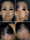
Parry-romberg syndrome with en coup de sabre
- PMID: 21909205
- PMCID: PMC3162264
- DOI: 10.5021/ad.2011.23.3.342
Parry-romberg syndrome with en coup de sabre
Abstract
Parry-Romberg syndrome (PRS) is a relatively rare degenerative disorder that is poorly understood. PRS is characterized by slowly progressing atrophy affecting one side of the face, and is frequently associated with localized scleroderma, especially linear scleroderma, which is known as en coup de sabre. This is a report of the author's experiences with PRS accompanying en coup de sabre, and a review of the ongoing considerable debate associated with these two entities. Case 1 was a 37-year-old woman who had right hemifacial atrophy with unilateral en coup de sabre for seven years. Fat grafting to her atrophic lip had been conducted, and steroid injection had been performed on the indurated plaque of the forehead. Case 2 was a 29-year-old woman who had suffered from right hemifacial atrophy and bilateral en coup de sabre for 18 years. Surgical corrections such as scapular osteocutaneous flap and mandible/maxilla distraction showed unsatisfying results.
Keywords: Encoup de sabre; Hemifacial atrophy; Parry-Romberg syndrome.
Figures


Similar articles
-
Parry-Romberg syndrome associated with en coup de sabre in a patient from South Sudan - a rare entity from East Africa: a case report.J Med Case Rep. 2019 May 3;13(1):138. doi: 10.1186/s13256-019-2063-2. J Med Case Rep. 2019. PMID: 31046814 Free PMC article.
-
Parry Romberg syndrome with localized scleroderma: A case report.J Clin Exp Dent. 2014 Jul 1;6(3):e313-6. doi: 10.4317/jced.51409. eCollection 2014 Jul. J Clin Exp Dent. 2014. PMID: 25136439 Free PMC article.
-
En coup de sabre morphea and Parry-Romberg syndrome: a retrospective review of 54 patients.J Am Acad Dermatol. 2007 Feb;56(2):257-63. doi: 10.1016/j.jaad.2006.10.959. Epub 2006 Dec 4. J Am Acad Dermatol. 2007. PMID: 17147965
-
Epilepsy in Parry-Romberg syndrome and linear scleroderma en coup de sabre: Case series and systematic review including 140 patients.Epilepsy Behav. 2021 Aug;121(Pt A):108068. doi: 10.1016/j.yebeh.2021.108068. Epub 2021 May 28. Epilepsy Behav. 2021. PMID: 34052630 Free PMC article.
-
A review of Parry-Romberg syndrome.J Am Acad Dermatol. 2012 Oct;67(4):769-84. doi: 10.1016/j.jaad.2012.01.019. Epub 2012 Mar 7. J Am Acad Dermatol. 2012. PMID: 22405645 Review.
Cited by
-
Disturbances of the stomatognathic system and possibilities of its correction in patients with craniofacial morphea.Postepy Dermatol Alergol. 2023 Oct;40(5):592-598. doi: 10.5114/ada.2023.131865. Epub 2023 Nov 9. Postepy Dermatol Alergol. 2023. PMID: 38028421 Free PMC article. Review.
-
Parry-Romberg syndrome affecting one half of the body.J Int Soc Prev Community Dent. 2016 Jul-Aug;6(4):387-90. doi: 10.4103/2231-0762.186792. J Int Soc Prev Community Dent. 2016. PMID: 27583230 Free PMC article.
-
Coexistence of Parry-Romberg syndrome with homolateral segmental vitiligo.Postepy Dermatol Alergol. 2013 Dec;30(6):409-11. doi: 10.5114/pdia.2013.39441. Epub 2013 Dec 18. Postepy Dermatol Alergol. 2013. PMID: 24494006 Free PMC article.
-
Parry Romberg syndrome: A case report and discussion.J Oral Maxillofac Pathol. 2012 Sep;16(3):406-10. doi: 10.4103/0973-029X.102498. J Oral Maxillofac Pathol. 2012. PMID: 23248475 Free PMC article.
-
Progressive hemifacial atrophy: a review.Orphanet J Rare Dis. 2015 Apr 1;10:39. doi: 10.1186/s13023-015-0250-9. Orphanet J Rare Dis. 2015. PMID: 25881068 Free PMC article. Review.
References
-
- Parry CH. Collections from the unpublished medical writings of the late Caleb Hillier Parry. London: Underwoods; pp. 1825–478.
-
- Romberg HM. Klinische Ergebnisse. Berlin: Forrtner; 1846. pp. 75–81.
-
- Taylor HM, Robinson R, Cox T. Progressive facial hemiatrophy: MRI appearances. Dev Med Child Neurol. 1997;39:484–486. - PubMed
-
- Goldberg-Stern H, deGrauw T, Passo M, Ball WS., Jr Parry-Romberg syndrome: follow-up imaging during suppressive therapy. Neuroradiology. 1997;39:873–876. - PubMed
-
- Jurkiewicz MJ, Nahai F. The use of free revascularized grafts in the amelioration of hemifacial atrophy. Plast Reconstr Surg. 1985;76:44–55. - PubMed
Publication types
LinkOut - more resources
Full Text Sources
Research Materials

