

A case of cutaneous bronchogenic cyst presenting with lymphoid follicles
- PMID: 21909217
- PMCID: PMC3162276
- DOI: 10.5021/ad.2011.23.3.392
A case of cutaneous bronchogenic cyst presenting with lymphoid follicles
Abstract
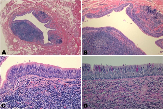
Cutaneous bronchogenic cysts are rare, and stem from developmental abnormalities of the tracheobronchial tree. The condition is often misdiagnosed clinically, with the correct diagnosis usually established by histopathologic examination. Published reports of bronchogenic or branchial anomalies are increasing, and the traditional defining characteristics of location and histopathology are proving to be less reliable for the identification of cutaneous bronchogenic cysts. In this report, we describe a case of a cutaneous bronchogenic cyst that presented with unusual histologic features, and was associated with several lymphoid follicles.
Keywords: Bronchogenic cyst; Lymphoid follicles.
Figures


Similar articles
-
Presternal bronchogenic sinus with predunculated lymphoid aggregate.Am J Dermatopathol. 2000 Feb;22(1):79-82. doi: 10.1097/00000372-200002000-00016. Am J Dermatopathol. 2000. PMID: 10698223
-
Cutaneous bronchogenic cyst: delineation of a poorly recognized lesion.Pediatr Dermatol. 1998 Jul-Aug;15(4):277-81. doi: 10.1046/j.1525-1470.1998.1998015277.x. Pediatr Dermatol. 1998. PMID: 9720691 Review.
-
A case report- retroperitoneal bronchogenic cyst in relation to the hindgut.Int J Surg Case Rep. 2020;75:140-142. doi: 10.1016/j.ijscr.2020.09.038. Epub 2020 Sep 12. Int J Surg Case Rep. 2020. PMID: 32950943 Free PMC article.
-
Multiple cutaneous bronchogenic cysts located on the neck and the scalp. A case report.Acta Dermatovenerol Alp Pannonica Adriat. 2008 Jun;17(2):69-71. Acta Dermatovenerol Alp Pannonica Adriat. 2008. PMID: 18709292
-
Esophageal bronchogenic cyst and review of the literature.Surg Endosc. 2015 Oct;29(10):3010-5. doi: 10.1007/s00464-015-4082-4. Epub 2015 Feb 11. Surg Endosc. 2015. PMID: 25669636 Review.
Cited by
-
Bronchogenic cysts with infection in the chest wall skin of a 64-year-old asymptomatic patient: A case report.World J Clin Cases. 2022 Aug 16;10(23):8392-8399. doi: 10.12998/wjcc.v10.i23.8392. World J Clin Cases. 2022. PMID: 36159540 Free PMC article.
References
-
- Fraga S, Helwig EB, Rosen SH. Bronchogenic cysts in the skin and subcutaneous tissue. Am J Clin Pathol. 1971;56:230–238. - PubMed
-
- Zvulunov A, Amichai B, Grunwald MH, Avinoach I, Halevy S. Cutaneous bronchogenic cyst: delineation of a poorly recognized lesion. Pediatr Dermatol. 1998;15:277–281. - PubMed
-
- Coleman WR, Homer RS, Kaplan RP. Branchial cleft heterotopia of the lower neck. J Cutan Pathol. 1989;16:353–358. - PubMed
-
- Miller OF, 3rd, Tyler W. Cutaneous bronchogenic cyst with papilloma and sinus presentation. J Am Acad Dermatol. 1984;11:367–371. - PubMed
-
- Søhoel P, Blom P, Mair IW. Subcutaneous bronchogenic anomalies. Ann Otol Rhinol Laryngol. 1980;89:75–77. - PubMed
Publication types
LinkOut - more resources
Full Text Sources
