

Case Reports
doi: 10.5021/ad.2011.23.3.400.
Epub 2011 Aug 6.
Acral Lentiginous Melanoma Developing during Long-standing Atypical Melanosis: Usefulness of Dermoscopy for Detection of Early Acral Melanoma
Affiliations
- PMID: 21909219
- PMCID: PMC3162278
- DOI: 10.5021/ad.2011.23.3.400
Item in Clipboard
Case Reports
Acral Lentiginous Melanoma Developing during Long-standing Atypical Melanosis: Usefulness of Dermoscopy for Detection of Early Acral Melanoma
Ann Dermatol.
2011 Aug.
Abstract
Clinical guidelines suggest that suspicious pigmented lesions of the plantar or palmar area require biopsy for early detection of acral melanoma. We present here a case of acral lentiginous melanoma in which various melanocytic atypia was observed at each biopsy site, including focal melanocytic proliferation. We suggest that this atypical melanosis is part of a contiguous phase of invasive tumor growth, which is known as the very early stage of melanoma in situ. In addition, noninvasive dermoscopy has been effective for the early discovery of hidden lesions of acral melanoma.
Keywords: Acral lentiginous melanoma; Dermoscopy.
Figures
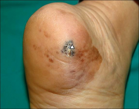
A 1.5×0.8-cm hyperkeratotic patch with irregular border and variegated color on the left heel. The lesion was surrounded by a sharply demarcated brownish patch with some mottled dark pigmentation. Initial punch biopsy site (white arrow).
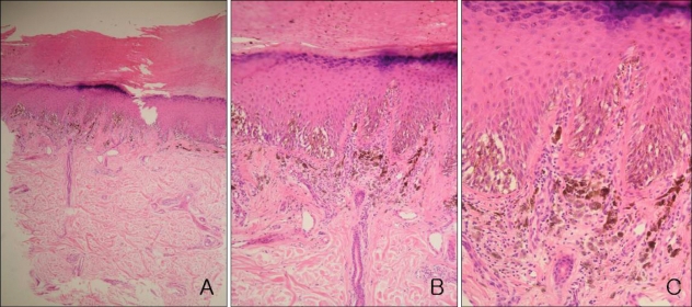
Increase in basal melanocytes and hyperpigmentation with focally uniform, severe cytologic atypia of melanocytes (A: H&E, ×40, B: H&E, ×100, C: H&E, ×200).

Typical parallel ridge pattern and abrupt cut-off of pigmentation observed upon dermoscopy.
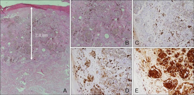
(A, B) Atypical melanocytes are nested at the upper dermis, with a Breslow thickness of 2.0 mm (A: H&E, ×40, B: H&E, ×200), (C) Melan A(-) (H&E, ×200), (D) HMB45(+, focal) (H&E, ×200), (E) S-100(+) (H&E, ×200).
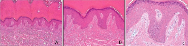
Histologic findings of peripheral brownish patch area, melanocytic proliferation predominantly in the crista profunda intermedia and diffuse basal hyperpigmenation (A: H&E, ×40, B: H&E, ×100, C: H&E, ×200).
Similar articles
-
Distinctive dermatoscopic features of acral lentiginous melanoma in situ from plantar melanocytic nevi and their histopathologic correlation.J Cutan Med Surg. 1998 Apr;2(4):199-204. doi: 10.1177/120347549800200404. J Cutan Med Surg. 1998. PMID: 9558302
-
Atypical melanosis of the foot. A report of three cases in Japanese populations.Arch Dermatol. 1994 Aug;130(8):1042-5. Arch Dermatol. 1994. PMID: 8053702
-
Acquired agminated melanocytic nevus in the acral area is a potential mimicker of acral lentiginous melanoma: A three-case series report and published work review.J Dermatol. 2020 Jul;47(7):770-773. doi: 10.1111/1346-8138.15353. Epub 2020 May 4. J Dermatol. 2020. PMID: 32363624
-
Role of In Vivo Reflectance Confocal Microscopy in the Analysis of Melanocytic Lesions.Acta Dermatovenerol Croat. 2018 Apr;26(1):64-67. Acta Dermatovenerol Croat. 2018. PMID: 29782304 Review.
-
Dermoscopic patterns of acral melanocytic lesions in skin of color.Cutis. 2019 May;103(5):274-276. Cutis. 2019. PMID: 31233579 Review.
Cited by
-
Recent advancements in the diagnosis and treatment of acral melanoma.J Zhejiang Univ Sci B. 2024 Feb 15;25(2):106-122. doi: 10.1631/jzus.B2300221. J Zhejiang Univ Sci B. 2024. PMID: 38303495 Free PMC article. Review.
-
Acral lentiginous melanoma in situ: dermoscopic features and management strategy.Sci Rep. 2020 Nov 25;10(1):20503. doi: 10.1038/s41598-020-77425-z. Sci Rep. 2020. PMID: 33239715 Free PMC article.
-
Progressive Acral Lentiginous Melanoma diagnosed via histopathology and surgically eradicated in a fingernail in a 69-year-old male - A Case Report.Int J Surg Case Rep. 2022 Sep;98:107611. doi: 10.1016/j.ijscr.2022.107611. Epub 2022 Sep 7. Int J Surg Case Rep. 2022. PMID: 36380543 Free PMC article.
-
Improving the diagnosis and treatment of acral melanocytic lesions.Melanoma Manag. 2017 May;4(2):113-123. doi: 10.2217/mmt-2016-0017. Epub 2017 May 19. Melanoma Manag. 2017. PMID: 30190914 Free PMC article. Review.
References
-
- Chiu HH, Hu SC, Ke CL, Cheng ST. Dermoscopy identifies histopathologically indiscernible malignant lesion of atypical melanosis of the foot, an early lesion of acral lentiginous melanoma in situ. Dermatol Surg. 2008;34:979–983. - PubMed
-
- Paek SC, Sober AJ, Tsao H, Mihm MC, Jr, Johnson TM. Cutaneous melanoma. In: Wolff K, Goldsmith LA, Katz SI, Gilchrest BA, Paller AS, Leffell DJ, editors. Fitzpatrick's dermatology in general medicine. 7th ed. New York: McGraw-Hill; 2008. pp. 1134–1157.
-
- Metzger S, Ellwanger U, Stroebel W, Schiebel U, Rassner G, Fierlbeck G. Extent and consequences of physician delay in the diagnosis of acral melanoma. Melanoma Res. 1998;8:181–186. - PubMed
-
- Somach SC, Taira JW, Pitha JV, Everett MA. Pigmented lesions in actinically damaged skin. Histopathologic comparison of biopsy and excisional specimens. Arch Dermatol. 1996;132:1297–1302. - PubMed
-
- Mishima Y, Nakanishi T. Acral lentiginous melanoma and its precursor--heterogeneity of palmo-plantar melanomas. Pathology. 1985;17:258–265. - PubMed
Publication types
LinkOut - more resources
Full Text Sources
