. 2011 Sep;7(9):e1002187.
doi: 10.1371/journal.ppat.1002187. Epub 2011 Sep 1.

Influenza virus ribonucleoprotein complexes gain preferential access to cellular export machinery through chromatin targeting

Affiliations

Influenza virus ribonucleoprotein complexes gain preferential access to cellular export machinery through chromatin targeting

Geoffrey P Chase et al. PLoS Pathog. 2011 Sep.

Abstract

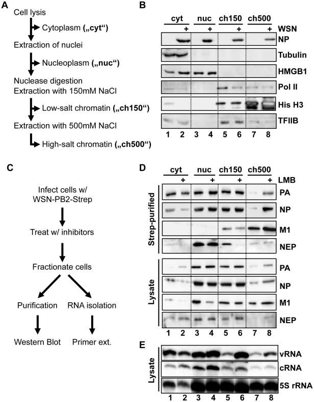
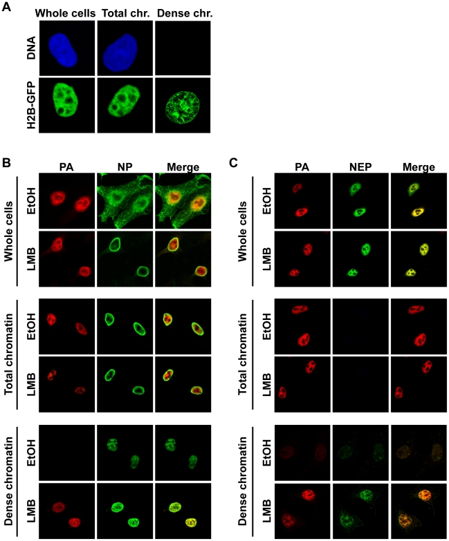
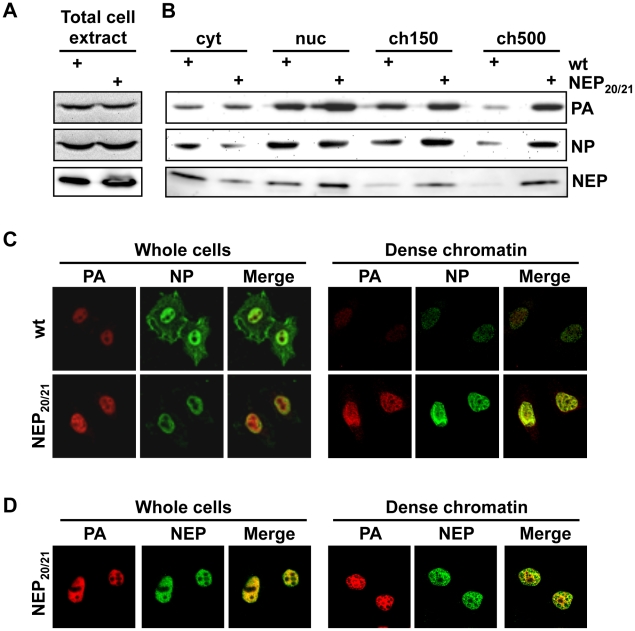
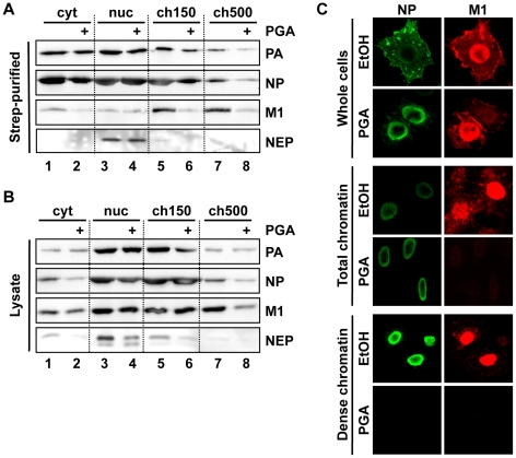
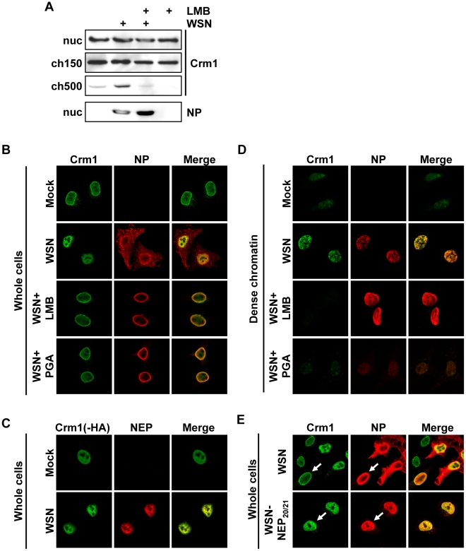
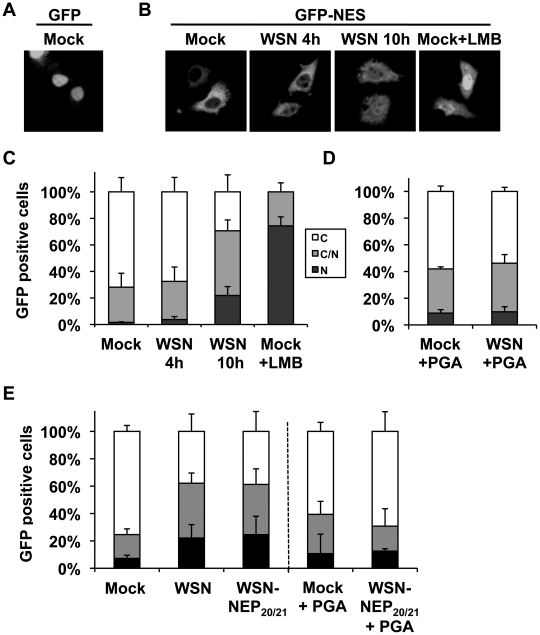
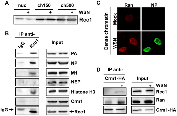
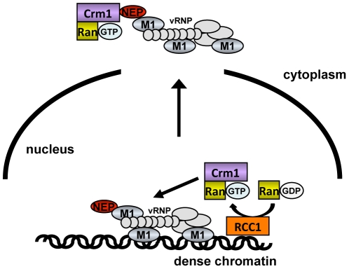
In contrast to most RNA viruses, influenza viruses replicate their genome in the nucleus of infected cells. As a result, newly-synthesized vRNA genomes, in the form of viral ribonucleoprotein complexes (vRNPs), must be exported to the cytoplasm for productive infection. To characterize the composition of vRNP export complexes and their interplay with the nucleus of infected cells, we affinity-purified tagged vRNPs from biochemically fractionated infected nuclei. After treatment of infected cells with leptomycin B, a potent inhibitor of Crm1-mediated export, we isolated vRNP export complexes which, unexpectedly, were tethered to the host-cell chromatin with very high affinity. At late time points of infection, the cellular export receptor Crm1 also accumulated at the same regions of the chromatin as vRNPs, which led to a decrease in the export of other nuclear Crm1 substrates from the nucleus. Interestingly, chromatin targeting of vRNP export complexes brought them into association with Rcc1, the Ran guanine exchange factor responsible for generating RanGTP and driving Crm1-dependent nuclear export. Thus, influenza viruses gain preferential access to newly-generated host cell export machinery by targeting vRNP export complexes at the sites of Ran regeneration.


Conflict of interest statement

The authors have declared that no competing interests exist.

Figures

Figure 1
Figure 1. Purification of vRNPs after subcellular fractionation.
(A) Schematic diagram of subcellular fractionation as described in Materials and Methods. (B) 1×10⁹ HeLa cells were infected with WSN at an MOI of 3 for 9 h or mock-infected. Cells were fractionated as described in (A), and equal amounts of protein from each fraction were analyzed by Western blot analysis for NP or cellular marker proteins. (C) Schematic workflow of Strep purification after LMB treatment. (D) 1×10⁹ HeLa cells were infected with rWSN-Strep at an MOI of 3 for 9 h. At 3hpi, cells were treated with 10 nM LMB or 0.2% EtOH vehicle. Fractionation and Strep-purification were performed as described in (C), and lysates and eluates were analyzed by Western blot for indicated proteins. (E) Cells were infected, treated, and fractionated as in (D), except that 50 U/ml DNase I was used as a nuclease, and total RNA was extracted from each fraction. RNA was analyzed by primer extension using probes specific for segment 6 v/cRNA and 5S rRNA.
Figure 2
Figure 2. Association of vRNP export complexes with subchromatin structure.
(A) The fractionation protocol was adapted to cells grown on coverslips. Cells were fixed after different fractionation steps to reveal the localization of unextracted proteins. HeLa cells were transfected with histone H2B-GFP or stained with ToPro3 after fixation to demonstrate extraction efficiency. Whole cells  =  no extraction, Total chr.  =  detergent extraction (all soluble proteins removed), Dense chr.  =  low-salt-extractable chromatin removed. (B) HeLa cells were grown on coverslips, infected with WSN at an MOI of 3 for 9 h, and treated with 10 nM LMB or 0.2% EtOH at 3 hpi. Cells were fractionated on the coverslips, fixed after fractionated, and stained for immunofluorescence using antibodies against PA and NP. Optical sections were obtained by confocal microscopy, and detector settings remained constant for each protein between all samples. (C) As in (B), except using antibodies against PA and NEP.
Figure 3
Figure 3. Accumulation of vRNPs on chromatin during infection with WSN-NEP20/21.
(A) HeLa cells were infected with WSN or WSN-NEP20/21 at an MOI of 3 for 7 h. Whole-cell lysates were analyzed by Western blot for accumulation of PA or NP. (B) 1×10⁹ HeLa cells were infected with WSN or WSN-NEP20/21 at an MOI of 3 for 9 h, then fractionated as in Fig. 1B. Fractionated lysates were analyzed by Western blot for PA, NP or NEP accumulation. (C) HeLa cells on coverslips were infected with WSN or WSN-NEP20/21 at an MOI of 3 for 9 h, then fractionated as described in Fig. 2B and stained for IFA using antibodies against PA and NP. (D) HeLa cells on coverslips were infected with WSN-NEP20/21 at an MOI of 3 for 9 h, then fractionated as described in Fig. 2A and stained for IFA using antibodies against PA and NEP.
Figure 4
Figure 4. Depletion of chromatin-bound vRNPs after PGA treatment.
(A) Cells were infected and purified as in Fig. 3A, except that treatment was performed using 20 µg/ml PGA at 3 hpi. Eluted complexes were analyzed by Western blot. (B) Lysates from (A) were analyzed by Western blot. (C) HeLa cells grown on coverslips were infected as in Fig. 4B, treated at 3 hpi with 20 µg/ml PGA, fractionated at 9 hpi as in Fig. 2A, and analyzed by immunofluorescence using antibodies specific for NP and M1. Note that the detector sensitivity was increased for the dense chromatin panels to demonstrate the absence of NP and M1 after PGA treatment.
Figure 5
Figure 5. Relocalization of Crm1 to dense chromatin after influenza virus infection.
(A) 1×10⁹ HeLa cells were infected with WSN at an MOI of 3 or mock-infected. At 3hpi, cells were treated with LMB or EtOH, and at 9hpi cells were fractionated. Lysates from the ch500 fraction were analyzed by Western blot for Crm1 or NP. (B) HeLa cells grown on coverslips were infected with WSN at an MOI of 3 or mock-infected. Cells were treated with EtOH, LMB or PGA at 3hpi, fixed at 9hpi, and analyzed by immunofluorescence using antibodies against NP and Crm1. (C) HeLa cells were transfected with Crm1-HA, infected with WSN at an MOI of 3 or mock-infected for 9 h, and analyzed by IFA using antibodies against NEP or HA. (D) Cells were infected and treated as in (B), then fractionated to reveal dense chromatin before fixation and immunofluorescence. (E) HeLa cells were infected with WSN or WSN-NEP20/21 for 9 h, and analyzed by IFA using antibodies against NP or Crm1.
Figure 6
Figure 6. Influenza infection impairs nuclear export of a Crm1-dependent protein.
(A) HeLa cells grown on coverslips were transfected with GFP, mock infected, and fixed. (B) HeLa cells grown on coverslips were transfected with GFP-NES and either infected with WSN at an MOI of 3, mock-infected, or treated with LMB. At the times indicated, cells were fixed, and GFP was detected by confocal microscopy. (C) Cells from (B) were scored for the percentage of GFP-expressing cells which displayed either predominantly cytoplasmic (C), approximately equal cytoplasmic and nuclear (C/N), or predominantly nuclear (N) GFP distribution. Values are the average of three independent assays. (D) HeLa cells grown on coverslips were transfected with GFP-NES and either infected or mock-infected at an MOI of 3 for 10 h, and treated with PGA at 3hpi. At the times indicated, cells were fixed, and GFP localization was scored as in (C). (E) HeLa cells transfected with GFP-NES were infected with WSN or WSN-NEP20/21 at an MOI of 3 for 9 h, and some treated with PGA at 3hpi. GFP localization was scored as in (C).
Figure 7
Figure 7. Association of Rcc1 with vRNPs and Crm1.
(A) 1×10⁹ HeLa cells were infected with WSN at an MOI of 3 or mock-infected for 9 h. Cells were fractionated, and nuclear lysates were analyzed for distribution of Rcc1. (B) 1×10⁹ HeLa cells were infected with WSN at an MOI of 3 for 9 h. Nuclease-digested lysates were incubated with anti-Rcc1 antibodies or pre-immune serum (IgG) and precipitated with Protein G agarose. Immunoprecipitates and input lysates were analyzed by Western blot for the indicated proteins. (C) HeLa cells grown on coverslips were infected with WSN at an MOI of 3 or mock-infected. Whole cells were fixed at 9hpi, and analyzed by IFA using antibodies against NP and Ran. (D) 2×10⁸ HeLa cells were transfected with an expression plasmid encoding HA-tagged Crm1. 16 h post-transfection, cells were infected with WSN at an MOI of 3 or mock-infected for 9 h. Cells were fractionated, and chromatin lysates were immunoprecipitated using monoclonal HA antibodies. Lysates and precipitates were normalized for Crm1 amounts (to accurately judge amounts of co-precipitated Ran and Rcc1) and analyzed by Western blot for HA-tagged Crm1 or endogenous Rcc1.
Figure 8
Figure 8. Model of vRNP export.
vRNPs (light gray) complexed with M1 (blue) and NEP (red) associate with dense chromatin in proximity to Rcc1 (orange). During or immediately after generation of RanGTP (purple), the proximity of vRNPs to Rcc1 may allow vRNPs access to a RanGTP-Crm1 complex before it can diffuse to other sites in the nucleus.

Similar articles

Cited by

References

    1. Martin K, Helenius A. Nuclear transport of influenza virus ribonucleoproteins: the viral matrix protein (M1) promotes export and inhibits import. Cell. 1991;67:117–130. - PubMed
    1. Neumann G, Castrucci MR, Kawaoka Y. Nuclear import and export of influenza virus nucleoprotein. J Virol. 1997;71:9690–9700. - PMC - PubMed
    1. O'Neill RE, Talon J, Palese P. The influenza virus NEP (NS2 protein) mediates the nuclear export of viral ribonucleoproteins. EMBO J. 1998;17:288–296. - PMC - PubMed
    1. Bui M, Wills EG, Helenius A, Whittaker GR. Role of the influenza virus M1 protein in nuclear export of viral ribonucleoproteins. J Virol. 2000;74:1781–1786. - PMC - PubMed
    1. Elton D, Simpson-Holley M, Archer K, Medcalf L, Hallam R, et al. Interaction of the influenza virus nucleoprotein with the cellular CRM1-mediated nuclear export pathway. J Virol. 2001;75:408–419. - PMC - PubMed

Publication types

MeSH terms