. 2011 Sep;7(9):e1002189.
doi: 10.1371/journal.ppat.1002189. Epub 2011 Sep 1.

Alterations in the Aedes aegypti transcriptome during infection with West Nile, dengue and yellow fever viruses

Affiliations

Alterations in the Aedes aegypti transcriptome during infection with West Nile, dengue and yellow fever viruses

Tonya M Colpitts et al. PLoS Pathog. 2011 Sep.

Abstract

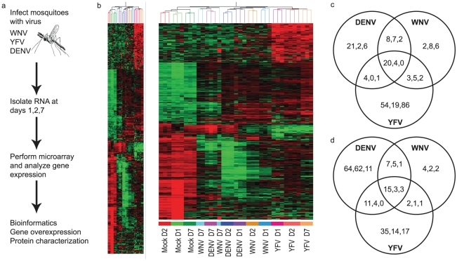
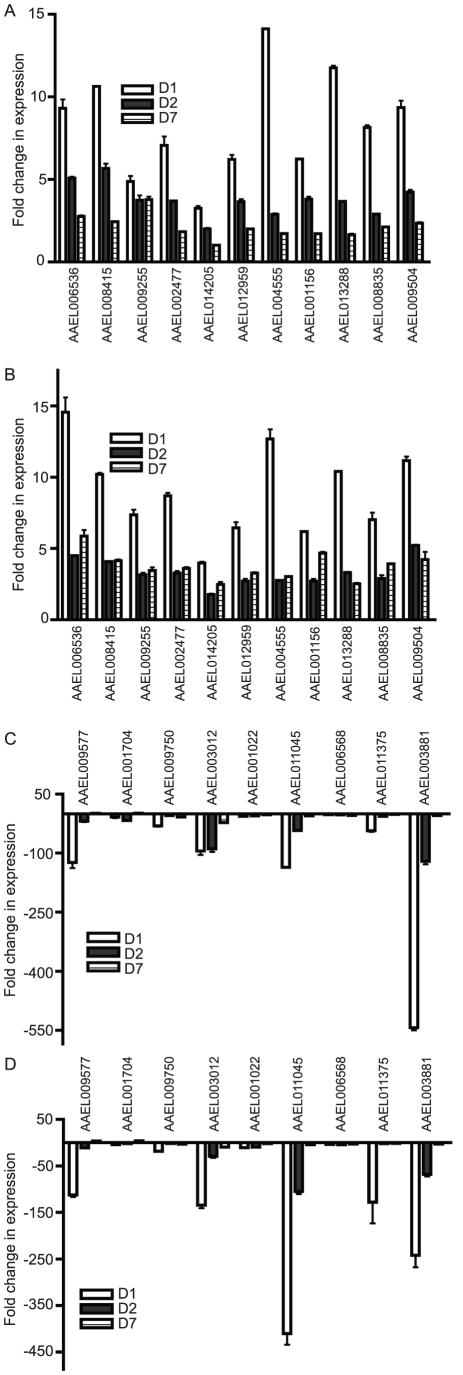
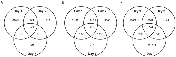
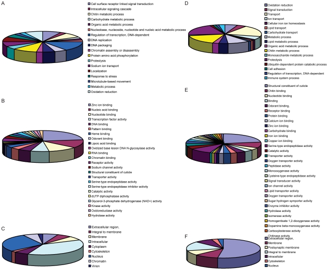
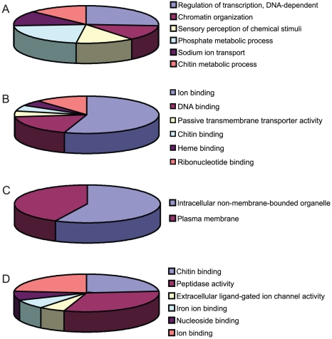
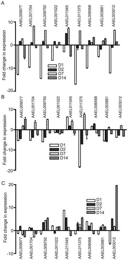
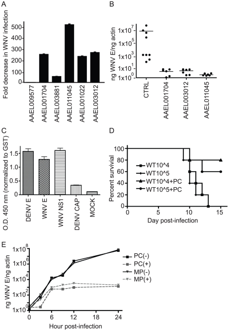
West Nile (WNV), dengue (DENV) and yellow fever (YFV) viruses are (re)emerging, mosquito-borne flaviviruses that cause human disease and mortality worldwide. Alterations in mosquito gene expression common and unique to individual flaviviral infections are poorly understood. Here, we present a microarray analysis of the Aedes aegypti transcriptome over time during infection with DENV, WNV or YFV. We identified 203 mosquito genes that were ≥ 5-fold differentially up-regulated (DUR) and 202 genes that were ≥ 10-fold differentially down-regulated (DDR) during infection with one of the three flaviviruses. Comparative analysis revealed that the expression profile of 20 DUR genes and 15 DDR genes was quite similar between the three flaviviruses on D1 of infection, indicating a potentially conserved transcriptomic signature of flaviviral infection. Bioinformatics analysis revealed changes in expression of genes from diverse cellular processes, including ion binding, transport, metabolic processes and peptidase activity. We also demonstrate that virally-regulated gene expression is tissue-specific. The overexpression of several virally down-regulated genes decreased WNV infection in mosquito cells and Aedes aegypti mosquitoes. Among these, a pupal cuticle protein was shown to bind WNV envelope protein, leading to inhibition of infection in vitro and the prevention of lethal WNV encephalitis in mice. This work provides an extensive list of targets for controlling flaviviral infection in mosquitoes that may also be used to develop broad preventative and therapeutic measures for multiple flaviviruses.


Conflict of interest statement

The authors have declared that no competing interests exist.

Figures

Figure 1
Figure 1. Genome-wide microarray analysis of the mosquito transcriptome during WNV, DENV and YFV infection.
A. Schematic of the experimental procedure. Ae.aegypti mosquitoes were infected with WNV, DENV or YFV. Microarray analysis was done at 1, 2 and 7 days p.i. for 15,959 genes. B. Heatmap for mosquito genes that were ≥5-fold up-regulated (203 genes) and ≥10-fold down-regulated (202 genes) during infection with any virus at any timepoint. Red, black and green colors indicate gene expression above, equal to and below the mean, respectively. C,D. Venn diagrams of the ≥5-fold up- (C) and ≥10-fold down-regulated (D) genes common and unique to each virus. Timepoints are indicated as D1/D2/D7 within each grouping. The complete dataset can be found in Table S1.
Figure 2
Figure 2. Expression of individual Ae. aegypti DEGs at 4 timepoints after WNV or DENV infection.
Mosquitoes were infected with virus via blood feeding and RNA isolated at 1, 2, 7 and 14 days post-infection. qRT-PCR analysis was done to measure expression of DURGs (A,B) and DDRGs (C,D) after WNV (A,C) or DENV (B,D) infection. Data from 3 separate infections were analyzed in triplicate and plotted on graphs, error bars indicate standard deviation. Fold change in expression was calculated from Ct values, normalized to actin and compared to mock infection.
Figure 3
Figure 3. Venn diagrams.
Venn diagrams of the ≥5-fold up- and ≥10-fold down-regulated genes common between timepoints for infection with WNV (A), DENV (B) and YFV (C). Up- and down-regulated genes are indicated as up/down within each grouping.
Figure 4
Figure 4. Functional annotations of DEGs.
DURGs (A–C) and DDRGs (D–F) were assigned to functional categories for biological process (A,D), molecular function (B,E) and cellular location (C,F). Genes with no known annotation are not included in the pie charts. The full list of annotations can be found in Table S2.
Figure 5
Figure 5. Functional clustering of DEGs.
DURGs (A–C) and DDRGs (D) were assigned to functional categories for biological process (A), molecular function (B,D) and cellular component (C). Genes with no known annotation are not included in the pie charts. The full list of functional clustering annotations can be found in Table S3.
Figure 6
Figure 6. Alteration in DDRG expression in Ae. aegypti is tissue-specific.
Gene expression was analyzed by qRT-PCR in mosquito tissues at select timepoints during WNV infection of Ae. aegypti mosquitoes. The fold change in expression of indicated genes in midgut (A), salivary gland (B) and abdomen (C), after WNV infection compared to mock infection. D1, D2, D7, D14 indicated by representative bars. Data is from 3 separate infection groups and qRT-PCR was done in triplicate. Error bars indicate standard deviation.
Figure 7
Figure 7. Overexpression of virally down-regulated genes impairs WNV infectivity.
A. Overexpression of DDRGs reduces WNV infection of Ae. aegypti mosquito cells. CCL-125 cells were transfected with expression plasmids encoding mosquito DDRGs and infected with WNV 48 hours post-transfection (p.t.). Cells were analyzed for infection by immunofluorescence microscopy 24 hours post-infection (p.i.) using an antibody against WNV envelope protein. Fold decrease in infection is indicated. Data is pooled from 3 separate experiments, error bars indicate standard deviation. B. Overexpression of DDRGs reduces WNV infection of Ae. aegypti mosquitoes. Mosquitoes were injected with plasmids used in (A) and infected with WNV 6 days p.t. At 10 days p.i., RNA was isolated and qRT-PCR performed to detect WNV. Infection is indicated as ng WNV E/ng actin. p<.001 for all 3 genes vs. control, one way ANOVA and Kruskal-Wallis test were used, n = 10. C. ELISA analysis of AAEL011045 (PC protein) binding to WNV E, DENV E, WNV NS1 and DENV capsid. Optical density (O.D.) is shown at 450 nm and normalized to glutathione S-transferase (GST) binding. Data is pooled from 3 separate experiments, error bars indicate standard deviation. D. Purified recombinant pupal cuticle protein (PC) (AAEL011045) was incubated with WNV for an hour at 37°C and injected i.p. into c57L/B6 mice. Survival curve was plotted for: 104 WNV alone, 105 WNV alone, 104 WNV+PC, 105 WNV+PC; number refers to viral titer in plaque forming units (pfu). p<.05 using the Logrank test. Results are pooled from 2 separate experiments, total n = 10 for each group. E. Overexpression of PC and MP protein affects WNV infection in mosquito cells. CCL-125 cells were transfected as in (A) and analyzed by qRT-PCR as in (B). Points plotted on line graph are from 3,6,9,12 and 24 h post-infection.

References

    1. Mackenzie JS, Gubler DJ, Petersen LR. Emerging flaviviruses: the spread and resurgence of Japanese encephalitis, West Nile and dengue viruses. Nat Med. 2004;10:S98–109. - PubMed
    1. Rappole JH, Derrickson SR, Hubalek Z. Migratory birds and spread of West Nile virus in the Western Hemisphere. Emerg Infect Dis. 2000;6:319–328. - PMC - PubMed
    1. Gubler DJ. Epidemic dengue/dengue hemorrhagic fever as a public health, social and economic problem in the 21st century. Trends Microbiol. 2002;10:100–103. - PubMed
    1. site PAHOw. 2007. Number of reported cases of dengue and dengue hemorrhagic fever (DHF) in the Americas, by country: figures for 2007.
    1. Blair CD, Adelman ZN, Olson KE. Molecular strategies for interrupting arthropod-borne virus transmission by mosquitoes. Clin Microbiol Rev. 2000;13:651–661. - PMC - PubMed

Publication types

MeSH terms

Substances