. 2011 Sep;7(9):e1002201.
doi: 10.1371/journal.ppat.1002201. Epub 2011 Sep 1.

The motility of a human parasite, Toxoplasma gondii, is regulated by a novel lysine methyltransferase

Affiliations

The motility of a human parasite, Toxoplasma gondii, is regulated by a novel lysine methyltransferase

Aoife T Heaslip et al. PLoS Pathog. 2011 Sep.

Abstract

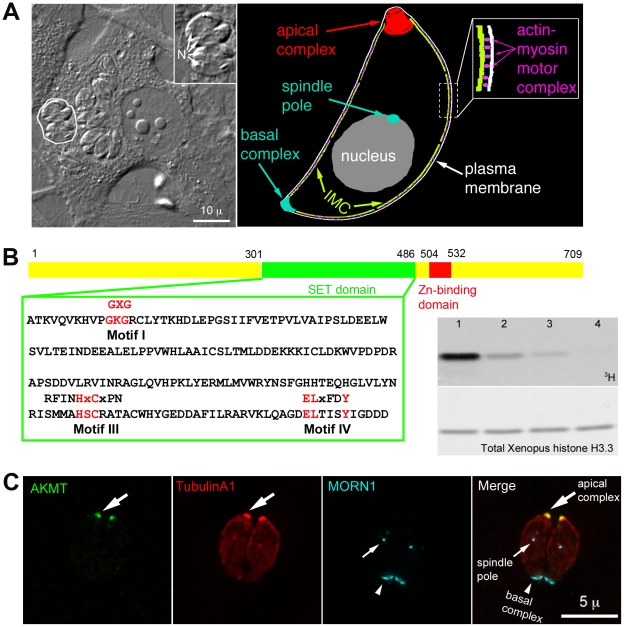
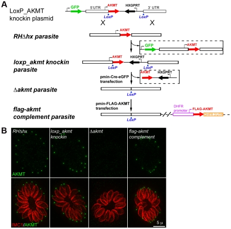
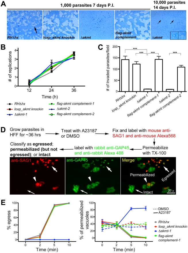
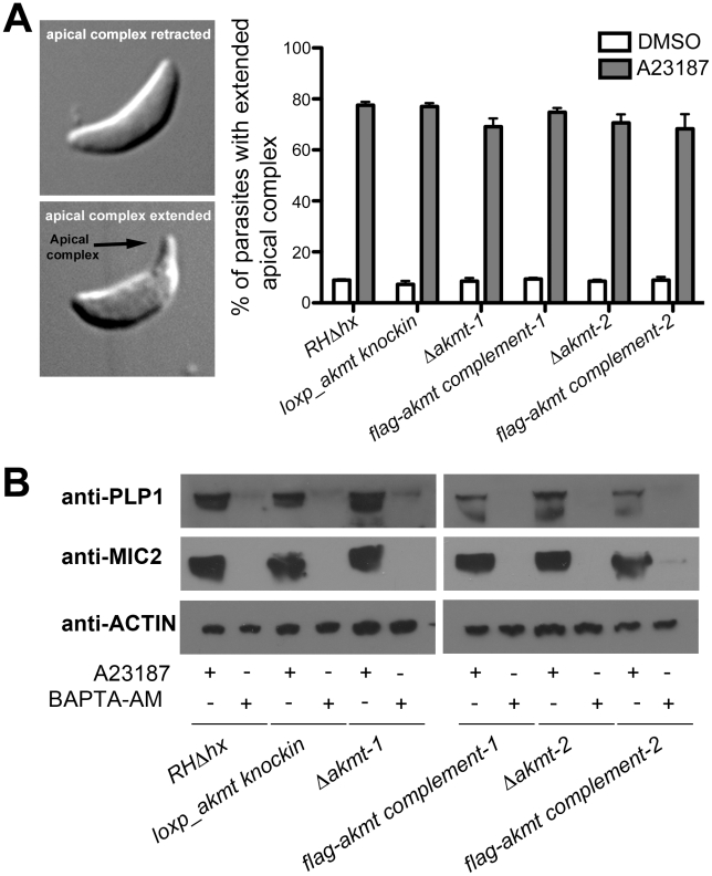
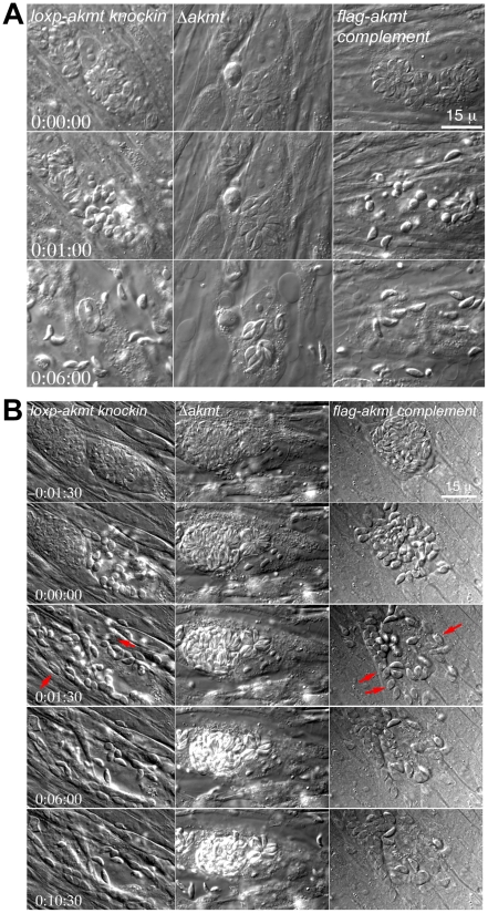
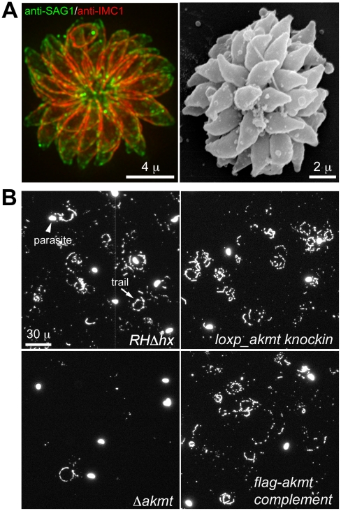
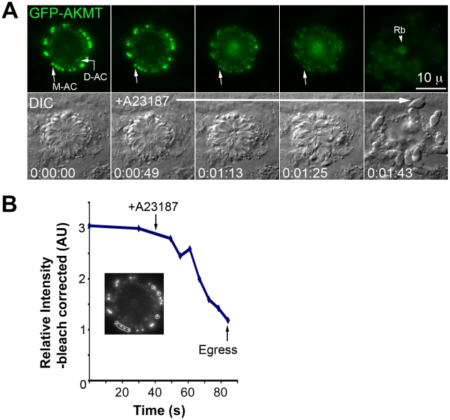
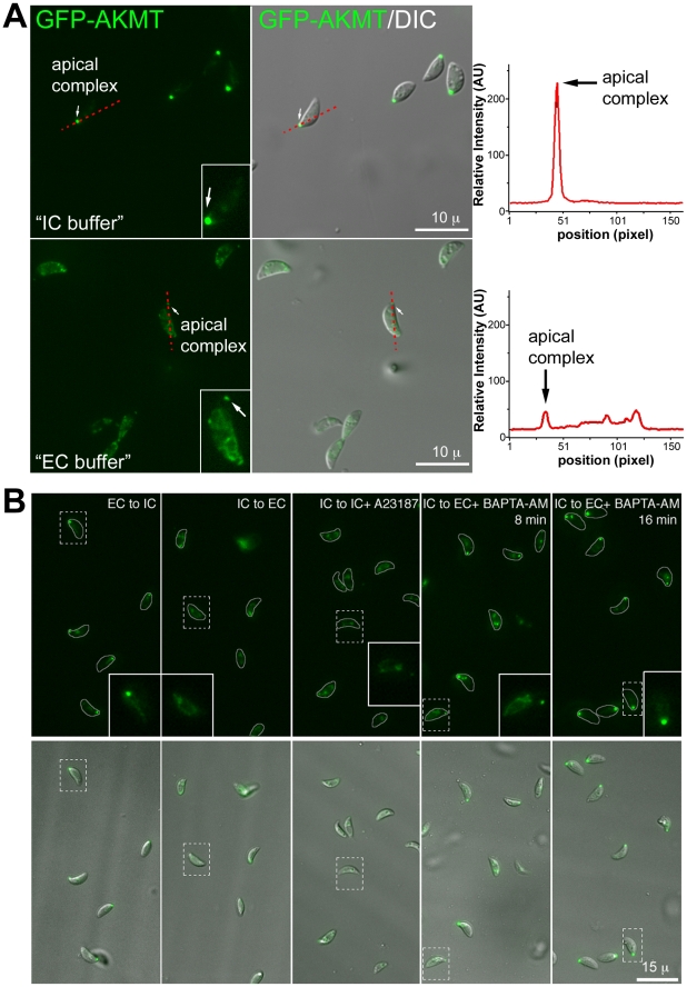
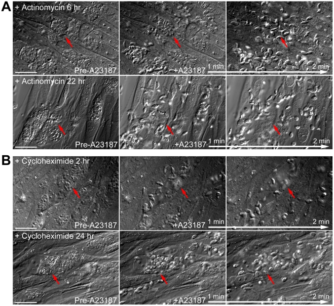
Protozoa in the phylum Apicomplexa are a large group of obligate intracellular parasites. Toxoplasma gondii and other apicomplexan parasites, such as Plasmodium falciparum, cause diseases by reiterating their lytic cycle, comprising host cell invasion, parasite replication, and parasite egress. The successful completion of the lytic cycle requires that the parasite senses changes in its environment and switches between the non-motile (for intracellular replication) and motile (for invasion and egress) states appropriately. Although the signaling pathway that regulates the motile state switch is critical to the pathogenesis of the diseases caused by these parasites, it is not well understood. Here we report a previously unknown mechanism of regulating the motility activation in Toxoplasma, mediated by a protein lysine methyltransferase, AKMT (for Apical complex lysine (K) methyltransferase). AKMT depletion greatly inhibits activation of motility, compromises parasite invasion and egress, and thus severely impairs the lytic cycle. Interestingly, AKMT redistributes from the apical complex to the parasite body rapidly in the presence of egress-stimulating signals that increase [Ca²⁺] in the parasite cytoplasm, suggesting that AKMT regulation of parasite motility might be accomplished by the precise temporal control of its localization in response to environmental changes.


Conflict of interest statement

The authors have declared that no competing interests exist.

Figures

Figure 1
Figure 1. AKMT is a novel lysine methyltransferase localized to the apical complex in intracellular parasites.
(A) Left: DIC image of human fibroblasts infected with T. gondii. The parasites live within a specialized parasitophorous vacuole, which is created from host cell plasma membrane during invasion. A parasitophorous vacuole containing 4 parasites (outline traced by a white border) is shown in the enlarged inset (N: nucleus). Inset: 1.5X magnification. Right: Cartoon drawing showing several membrane and cytoskeletal structures referred to in the text. For clarity, cortical microtubules of the parasite are not shown. Also not shown is the portion of the motility apparatus that mediates the interaction between the parasite and the host cell surface. (B) Left: AKMT contains SET (green) and zinc-binding (red) domains and is a predicted PKMT. The SET domain of canonical PKMTs contain four highly conserved motifs that are involved in different aspects of methyltransferase reactions: motif I for SAM binding; motif II for catalytic methyl transfer; motif III and IV for SAM binding and target lysine-binding. Three of the four motifs found in canonical PKMTs can be identified in AKMT, which contains a well-conserved motif I, and semi-conserved motifs III and IV. However, no conservation with the conventional motif II can be found in AKMT. Conserved amino acids in each motif found in canonical SET domains are shown above the T. gondii AKMT sequence. Right: Despite the unconventional SET domain, AKMT is a functional KMT enzyme. It can methylate an artificial substrate, Xenopus histone H3.3, in vitro in the presence of 3H-S-adenyl-L-methionine (3H-SAM). Top: 3H signal of histone in PKMT reactions containing 3H-SAM; 1 µg Xenopus histone H3.3; and recombinant FLAG-AKMT in the amount of 0.5 µg (lane1), 0.1 µg (lane 2), 0.05 µg (lane 3), 0.01 µg (lane 4). Bottom: The same blot stained with amido black to show total histone H3.3. Histone labeling can be detected when as low as 0.05 µg (lane 3) of FLAG-AKMT is present in the reaction. (C) Fluorescence images of two intracellular parasites at interphase showing that AKMT is localized to the apical complex (large arrows), a pattern observed in 100% of the hundreds of interphase parasites examined. Green: anti-AKMT; red: mCherryFP-TubulinA1 , highlighting all the tubulin containing structure in the parasite, including the main body of the cytoskeletal apical complex (large arrows); cyan: eGFP-MORN1 , , highlighting the basal complex of the parasite (arrowheads) as well as the spindle pole (small arrows). The host cell is not visible in these images, because it is not fluorescent.
Figure 2
Figure 2. The generation and characterization of the AKMT knockout (Δakmt) mutant.
(A) Procedure for generating the Δakmt parasite line. See text for details. (B) Immunofluoresence assay of intracellular RHΔhx, loxp_akmt knockin, Δakmt and flag-akmt complement parasites showing undetectable levels of AKMT in Δakmt parasites and the correct localization of FLAG-AKMT in the complemented parasites. Green: anti-AKMT; red: anti-IMC1. Each image shows 16 parasites contained within one parasitophorous vacuole. The host cells are not visible in these images, because they are not fluorescent.
Figure 3
Figure 3. AKMT is important for invasion and egress, but not for parasite replication.
(A) The lytic cycle of Δakmt parasites is greatly compromised. Left: Images showing portions of T12.5 tissue culture flasks containing a monolayer of HFF cells inoculated with 1000 parasites (RHΔhx, loxp_akmt knockin, Δakmt or flag-akmt complement parasites), grown for 7 days. The samples were then fixed and stained with Commassie Blue. The cultures were stained blue except for “plaques” (arrow) in which host cells had been destroyed by many rounds of parasite lytic cycles. No plaques were seen in the culture infected with Δakmt parasites. Right: HFF culture infected with 10,000 Δakmt parasites for 14 days. Only two small plaques (small arrows, insets) were detected. Insets 2X magnification. P.I.: Post Infection. (B) The loss of AKMT does not affect parasite replication. Parasites were grown for 12, 24 or 36 hrs and the number of parasites/vacuole was counted in 100 vacuoles/experiment. The average number of replications that had occurred at each time point was calculated from the results of three independent experiments. (C) The loss of AKMT results in a ∼90% decrease in invasion efficiency. The number of invaded parasites per field was counted in 10 fields each in 3 independent experiments. ***: P value <0.0001 (D) Top: Outline of A23187 induced egress assay. Bottom: Fluorescence images showing examples of three classes of parasitophorous vacuoles scored in the induced egress assay: 1) egressed if parasites had dispersed from the vacuole (large arrows); 2) permeabilized if parasites were retained in the parasitophorous vacuole but some or all of the parasites in the vacuole were labeled with anti-SAG1 antibody (arrowheads); 3) intact if none of the parasites in the vacuole were labeled with anti-SAG1 antibody (small arrows). (E) The loss of AKMT results in severe impairment in A23187 induced parasite egress despite efficient host cell permeabilization. Left: Percentage of vacuoles where egress had occurred 0, 2, 5, and 10 minutes after A23187 treatment. Right: Percentage of vacuoles that had been permeabilized 0, 2, 5, and 10 minutes after DMSO (dashed lines) or 5 µM A23187 (solid lines) treatment. Note that the AKMT expressing parasites (RHΔhx, loxp_akmt knockin, or flag-akmt complement) treated with A23817 have an apparent low percentage of permeabilized vacuoles at late time points (5 and 10 minutes), because most of the parasites have already egressed (see left). Total 50 vacuoles at each time point were scored for each of the three independent experiments. The behaviors of the two Δakmt lines were very similar, and both lines were fully complemented by FLAG-AKMT expression. For clarity, only the results from Δakmt-1 and the corresponding complemented line, flag-akmt complement-1 were included in the graph.
Figure 4
Figure 4. Ca2+ influx induced apical complex extension and microneme secretion are not affected in extracellular Δakmt parasites.
(A) A23187 stimulated apical complex extension is not affected in extracellular Δakmt parasites. Left: DIC images of T. gondii with the cytoskeletal apical complex (arrow) retracted (top) or extended (bottom). Right: Percentage of parasites with extended apical complex when treated with DMSO (white bars) or 5 µM A23187 (gray bars). 300 parasites/slide were scored for apical complex extension in each of three independent experiments. (B) Microneme secretion assays analyzed by Western blot showing that the A23187 stimulated secretion of two microneme proteins, TgPLP1 (one of the proteins responsible for permeabilizing the host cell during parasite egress) and TgMIC2 (one of the adhesive components of the motility apparatus) , , , , , is not affected in extracellular Δakmt parasites. Treatment with BAPTA-AM, a cell-permeant calcium chelator that inhibits microneme secretion , was used as the negative control for the secretion assay. Actin was used as the loading control.
Figure 5
Figure 5. AKMT regulates parasite motility activation.
(A) Images selected from time-lapse experiments of intracellular loxp_akmt knockin, Δakmt and flag-akmt complement parasites treated with 5 µM A23187 (also see Video S1). (B) Images selected from natural egress time-lapse experiments of intracellular loxp_akmt knockin, Δakmt and flag-akmt complement parasites (also see Video S2). To display the egress process in synchrony, the time point at which parasite egress (for loxp_akmt knockin and flag-akmt complement parasites) or host cell permeabilization (for Δakmt parasites) occurs is chosen as the 0∶00∶00 time point for all videos. Notice that for loxp_akmt knockin and flag-akmt complement parasites, host cell permeabilization and parasite egress occur almost simultaneously and many parasites (red arrows) invade adjacent host cells immediately after egress. Δakmt parasites however, remain largely immotile and fail to exit the host cell actively.
Figure 6
Figure 6. The motility of Δakmt parasites is impaired.
(A) “Extracellular rosettes” found in Δakmt parasite culture. Left: Fluorescence image of a Δakmt extracellular rosette, a cluster of parasites remaining attached to each other after host cell breakdown. Green: anti-SAG1; red: anti-IMC1. Right: Scanning electron microscopy image of a Δakmt extracellular rosette. (B) Trail assays of RHΔhx, loxp_akmt knockin, Δakmt and flag-akmt complement parasites, showing that there is a marked decrease in trail deposition in Δakmt parasites compared to the parental and complemented lines. Mouse anti-SAG1 antibody and goat anti-mouse Alexa488 were used to visualize trails (arrow) deposited by the parasites.
Figure 7
Figure 7. The lysine methyltransferase activity of AKMT is required for its function.
(A) The mutation H447V abolished the enzyme activity of AKMT in vitro. Left: Blot stained with amido black to show total protein of histone H3.3+3H-SAM (lane 1); histone H3.3+1 µg FLAG-AKMT(WT)+3H-SAM (lane 2); and histone H3.3+1 µg FLAG-akmt(H447V)+3H-SAM (lane 3). Right: 3H signal of the same blot. Arrowheads indicate the positions of AKMT and histone H3 on the blots. Note that in the autoradiograph of FLAG-AKMT(WT) PKMT reaction (lane2), there is a weak band corresponding to AKMT self-methylation. (B) eGFP-akmt(H447V) failed to complement the egress defect of Δakmt parasites. Images show the results from an induced egress time-lapse experiment of intracellular Δakmt parasites expressing eGFP-AKMT(WT) (top) or eGFP-akmt(H447V) (bottom) parasites treated with 5 µM A23187. Arrows: apical complex of mature parasites. Arrowhead: daughter apical complex.
Figure 8
Figure 8. Elevated [Ca2+] triggers the dispersal of AKMT from the apical complex before A23187 induced parasite egress.
(A) Images show the result from a time-lapse experiment of intracellular Δakmt parasites expressing eGFP-AKMT(WT) treated with 5 µM A23187 (A23187 was added between 0∶00∶30 and 0∶00∶49). These parasites were in the process of daughter construction as indicated by the two internal AKMT positive spots (daughter apical complexes: D-AC) in addition to the AKMT labeling in the mother apical complex (M-AC) of each parasite (c.f. Figure S1C). These parasites are connected through a structure in the central region called the residual body (Rb, arrowhead) . Upon treatment with A23187, eGFP-AKMT relocated from the mother apical complex to the parasite body, followed by parasite egress. The residual body, containing some of the eGFP-AKMT released from the apical complex, is left behind. (B) Plot of average intensity (bleach-corrected) of eGFP-AKMT over time for seven mother apical complexes (encircled in the gray-scale image) in the experiment shown in A, showing the dissociation of AKMT from the mother apical complex before parasite egress. AU: Arbitrary Unit.
Figure 9
Figure 9. The localization of AKMT is affected by the extracellular and intracellular ionic composition.
(A) Left: Images show the localization of AKMT in extracellular egfp-akmt complement parasites incubated either in a potassium-based buffer mimicking intracellular conditions (“IC buffer”, top) or a sodium-based buffer mimicking extracellular conditions (“EC buffer”, bottom). Inset 2X magnification. Right: Intensity plots along the red dashed lines in the images shown on the left. AU: Arbitrary Unit. (B) Images from buffer exchange experiments where extracellular egfp-akmt complement parasites were first incubated in EC or IC buffer for 25–30 minutes, and then were resuspended in buffer containing different ionic composition or drugs that affect intra-parasite [Ca2+] concentration. From left to right showing: “EC to IC”- image of egfp-akmt complement parasites ∼32 minutes after buffer exchange from EC to IC buffer, which resulted in redistribution of AKMT from parasite body to the apical complex, a process that took ∼30 minutes to complete; “IC to EC”- image of egfp-akmt complement parasites ∼7 minutes after buffer exchange from IC to EC buffer, showing eGFP-AKMT dissociated from the apical complex; “IC to IC+A23187”- image of egfp-akmt complement parasites ∼5 minutes after buffer exchange from IC to IC buffer +5 µM A23187, showing eGFP-AKMT dissociated from the apical complex; “IC to EC+BAPTA-AM”- images of egfp-akmt complement parasites 8 and 16 minutes after buffer exchange from IC buffer to EC buffer+50 µM BAPTA-AM, showing a gradual relocation of AKMT from the parasite body to the apical complex. This change was noticeable by 7–8 minutes, and completed by ∼15–16 minutes after the buffer exchange. Top: fluorescence images super-imposed with the outlines of the parasites (gray borders). Bottom: overlay images of fluorescence and DIC. Inset 2X magnification
Figure 10
Figure 10. New RNA transcription and protein synthesis is not required for motility activation in induced egress.
(A) Images show the results from time-lapse experiments of RHΔhx parasites treated with 0.5 µg/ml actinomycin D (an inhibitor of RNA transcription) for 6 (top) or 22 hours (bottom) showing that this treatment has no effect on parasite motility stimulated by 5 µM A23817. Red arrows provide reference points for following changes of the parasitophorous vacuoles indicated in each sequence. Note the obvious lack of parasite growth in the presence of actinomycin D, comparing the size of the parasitophorous vacuoles in the culture treated for 6 versus 22 hours (Actinomycin D treatment for the two cultures was initiated simultaneously ∼26 hours post-infection.). Bars = 20 µm. (B) Images show the results from time-lapse experiments of RHΔhx parasites treated with 10 µM cycloheximide (an inhibitor of protein translation) for 2 (top) or 24 hours (bottom) showing that this treatment has no effect on parasite motility stimulated by 5 µM A23817. Red arrows provide reference points for following changes of the parasitophorous vacuoles indicated in each sequence. Bars = 20 µm.

Similar articles

Cited by

References

    1. Levine ND. Progress in taxonomy of the Apicomplexan protozoa. J Protozool. 1988;35:518–520. - PubMed
    1. Dobrowolski JM, Carruthers VB, Sibley LD. Participation of myosin in gliding motility and host cell invasion by Toxoplasma gondii. Mol Microbiol. 1997;26:163–173. - PubMed
    1. Lingelbach K, Joiner KA. The parasitophorous vacuole membrane surrounding Plasmodium and Toxoplasma: an unusual compartment in infected cells. J Cell Sci. 1998;111:1467–1475. - PubMed
    1. Sheffield HG, Melton ML. The fine structure and reproduction of Toxoplasma gondii. J Parasitol. 1968;54:209–226. - PubMed
    1. Sinai AP, Joiner KA. Safe haven: the cell biology of nonfusogenic pathogen vacuoles. Annu Rev Microbiol. 1997;51:415–462. - PubMed

Publication types