. 2011 Sep;7(9):e1002217.
doi: 10.1371/journal.ppat.1002217. Epub 2011 Sep 1.

c-di-AMP is a new second messenger in Staphylococcus aureus with a role in controlling cell size and envelope stress

Affiliations

c-di-AMP is a new second messenger in Staphylococcus aureus with a role in controlling cell size and envelope stress

Rebecca M Corrigan et al. PLoS Pathog. 2011 Sep.

Abstract

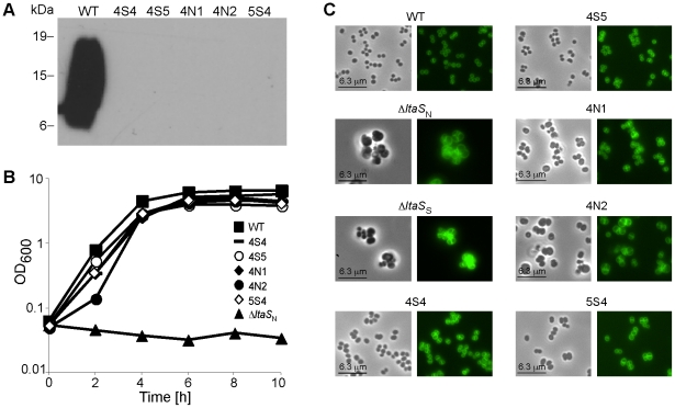
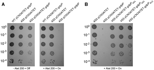
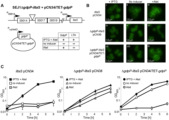
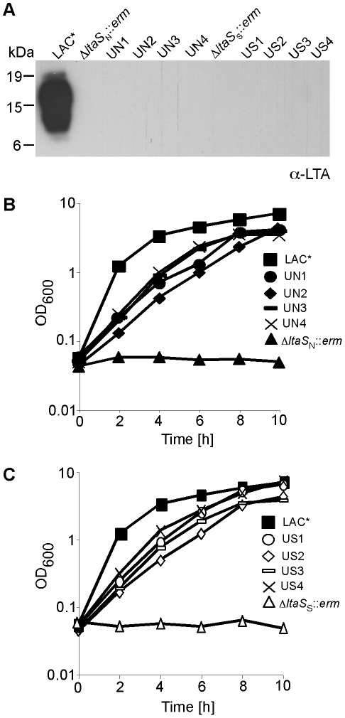
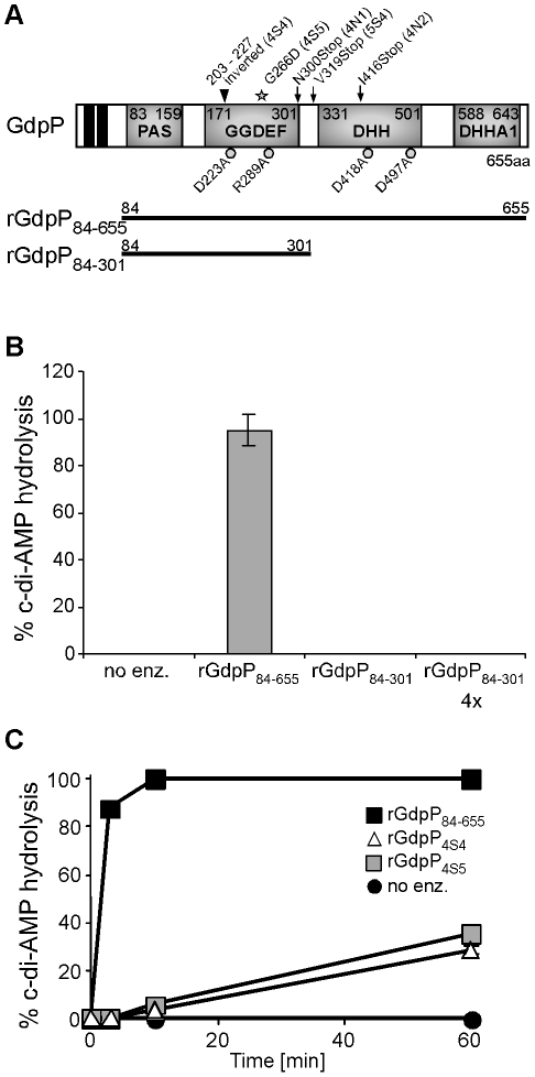
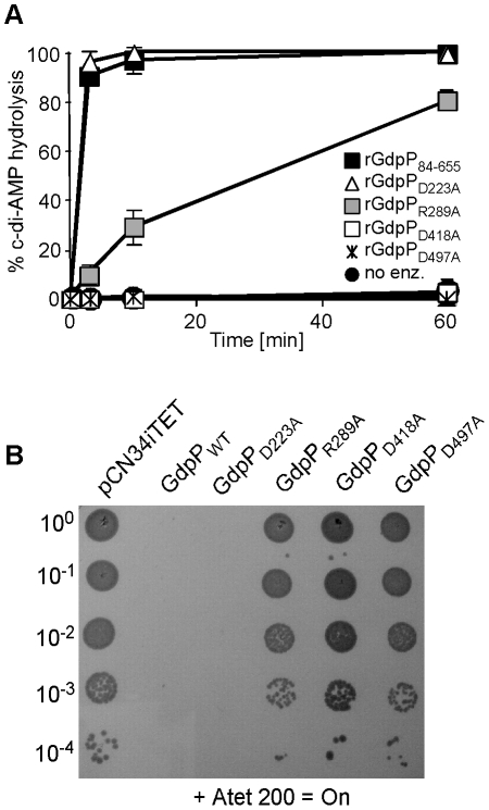
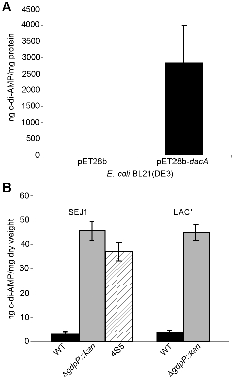
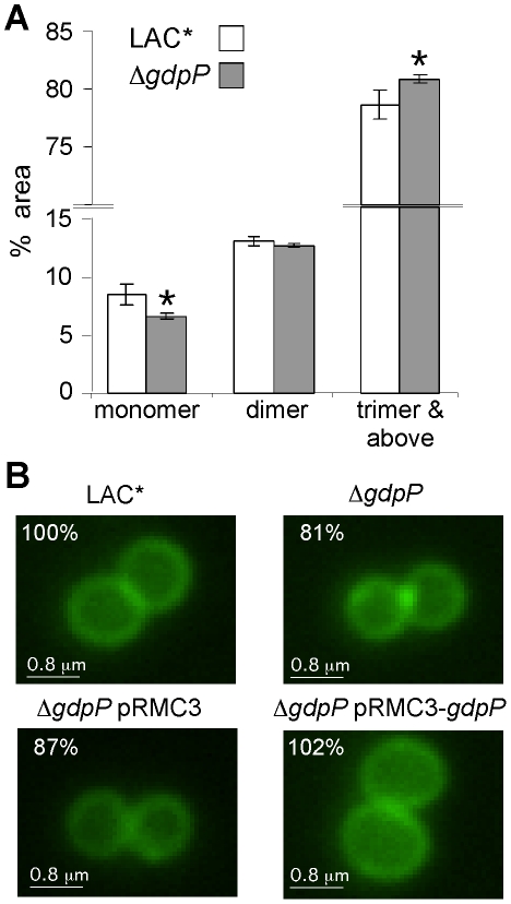
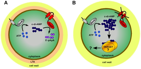
The cell wall is a vital and multi-functional part of bacterial cells. For Staphylococcus aureus, an important human bacterial pathogen, surface proteins and cell wall polymers are essential for adhesion, colonization and during the infection process. One such cell wall polymer, lipoteichoic acid (LTA), is crucial for normal bacterial growth and cell division. Upon depletion of this polymer bacteria increase in size and a misplacement of division septa and eventual cell lysis is observed. In this work, we describe the isolation and characterization of LTA-deficient S. aureus suppressor strains that regained the ability to grow almost normally in the absence of this cell wall polymer. Using a whole genome sequencing approach, compensatory mutations were identified and revealed that mutations within one gene, gdpP (GGDEF domain protein containing phosphodiesterase), allow both laboratory and clinical isolates of S. aureus to grow without LTA. It was determined that GdpP has phosphodiesterase activity in vitro and uses the cyclic dinucleotide c-di-AMP as a substrate. Furthermore, we show for the first time that c-di-AMP is produced in S. aureus presumably by the S. aureus DacA protein, which has diadenylate cyclase activity. We also demonstrate that GdpP functions in vivo as a c-di-AMP-specific phosphodiesterase, as intracellular c-di-AMP levels increase drastically in gdpP deletion strains and in an LTA-deficient suppressor strain. An increased amount of cross-linked peptidoglycan was observed in the gdpP mutant strain, a cell wall alteration that could help bacteria compensate for the lack of LTA. Lastly, microscopic analysis of wild-type and gdpP mutant strains revealed a 13-22% reduction in the cell size of bacteria with increased c-di-AMP levels. Taken together, these data suggest a function for this novel secondary messenger in controlling cell size of S. aureus and in helping bacteria to cope with extreme membrane and cell wall stress.


Conflict of interest statement

The authors have declared that no competing interests exist.

Figures

Figure 1
Figure 1. Characterization of ΔltaS suppressor strains.
(A) LTA detection by western blot. Cell-associated LTA isolated from strain SEJ1 (WT) and the five suppressor strains (4S4, 4S5, 4N1, 4N2 and 5S4) was analyzed by western blot. The positions of protein molecular mass markers (in kDa) are indicated on the left. (B) Bacterial growth curves. Overnight cultures of SEJ1 (WT) and the five suppressor strains (4S4, 4S5, 4N1, 4N2 and 5S4) were grown in TSB. Strain SEJ1ΔltaS NltaS N) was grown overnight in TSB 7.5% NaCl. All cultures were washed 3 times in TSB and diluted to a starting OD600 of 0.05 in TSB. Growth was monitored over a 10 h period. (C) Microscopic analysis of WT and LTA-negative strains. SEJ1 (WT) and the LTA-negative strains (4S4, 4S5, 4N1, 4N2 and 5S4) were grown in TSB to log-phase and strains SEJ1ΔltaS NltaS N) and SEJ1ΔltaS SltaS S) were grown in TSB containing 7.5% NaCl or 40% sucrose, respectively. Samples were prepared for microscopy analysis and bacteria stained with BODIPY-vancomycin as described in the materials and methods section.
Figure 2
Figure 2. Expression of different gdpP alleles in an LTA-negative suppressor strain and their effect on growth.
(A) Serial dilutions (indicated on the left) of WT SEJ1 containing the Atet-inducible GdpP expression vector (WT pCN34iTET-gdpP), the suppressor strain 4S5 containing the empty vector (4S5 pCN34iTET) or the GdpP expression vector (4S5 pCN34iTET-gdpP) were spotted on TSA plates containing either no (left panel) or 200 ng/ml Atet (right panel). (B) The mutated gdpP alleles from suppressor strains 4S4, 4S5 and 4N2 (indicated above panel) were expressed from pCN34iTET in the strain 4S5. Serial dilutions of were spotted on TSA 200 ng/ml Atet plates to induce protein expression from the plasmid vector.
Figure 3
Figure 3. Deletion of gdpP can rescue the dependence of RN4220iltaS on IPTG.
(A) Schematic representation of S. aureus strain SEJ1ΔgdpP-iltaS pCN34iTET-gdpP, containing an inframe gdpP deletion, the ltaS gene under IPTG inducible Pspac control and the complementing vector pCN34iTET-gdpP. The effects of the addition of the inducers on the production of LTA and GdpP are indicated. (B) Microscopic analysis of strains RN4220iltaS pCN34 (iltaS pCN34; i–iii), SEJ1ΔgdpP-iltaS pCN38 (ΔgdpP-iltaS pCN38; iv–vi) and SEJ1ΔgdpP-iltaS pCN34iTET-gdpPgdpP-iltaS pCN34iTET-gdpP; vii–ix). Strains were grown overnight in the presence of IPTG, washed and grown for 8 h in TSB containing 1mM IPTG (left panel), TSB (no inducer – middle panel) or TSB containing 100 ng/ml Atet (right panel). Bacteria were stained with BODIPY-vancomycin and prepared for microscopy analysis as described in the materials and methods section. Larger fields of view are shown in Figure S3B in Text S1 (C) Bacterial growth in the presence or absence of inducer. Strains RN4220iltaS pCN34 (iltaS pCN34), SEJ1ΔgdpP-iltaS pCN38 (ΔgdpP-iltaS pCN38) and SEJ1ΔgdpP-iltaS pCN34iTET-gdpPgdpP-iltaS pCN34iTET-gdpP) were grown overnight in the presence of IPTG, washed and diluted in either TSB containing 1mM IPTG +100 ng/ml Atet (black symbols), TSB containing 100 ng/ml Atet (grey symbols) or just plain TSB (no inducer – white symbols). After 4 h cultures were diluted 1:100 into fresh medium to maintain them in the exponential growth phase and this OD600 is plotted as T = 0. Growth was then continued for a further 6 h. Growth curves were performed in triplicate and the average and standard deviations were plotted.
Figure 4
Figure 4. Growth and LTA production of CA-MRSA LAC* and LAC*ΔltaS strains.
(A) LTA detection by western blot. S. aureus LAC* (WT) and the eight suppressor strains UN1, UN2, UN3, UN4, US1, US2, US3 and US4 were grown in TSB. LAC*ΔltaS N ::erm was grown in TSB containing 7.5% NaCl and LAC*ΔltaS S ::erm in TSB containing 40% sucrose and LTA detected by western blot. (B) and (C) Bacterial growth curves. Overnight cultures of strains described above were washed 3 times and diluted to a starting OD600 of 0.05 in TSB and OD600 readings determined every 2 h for 10 h.
Figure 5
Figure 5. In vitro phosphodiesterase activity of WT and mutant GdpP variants.
(A) Schematic representation of the GdpP protein. The N-terminus of the protein contains 2 transmembrane helices (black boxes), a degenerate PAS domain (residues 83–159), a GGDEF domain (residues 171–301), a DHH domain (residues 331–501) and a DHHA1 domain (residues 588–643). Protein domains expressed as recombinant proteins are indicated by the thick black lines. Amino acids mutated to alanines by site-directed mutagenesis are indicated by grey circles and residues altered in suppressor strains are indicated by a grey star (amino acid substitution – 4S5), by black arrows (stop codons – 4N1, 4N2 and 5S4) or by a triangle (inframe amino acid inversion – 4S4). (B) and (C) Phosphodiesterase activity of rGdpP proteins. (B) Enzyme reactions were set up in assay buffer containing 20 µM c-di-AMP and either no enzyme (no enz.), 1 µM rGdpP84–655, 1 µM rGdpP84–301 or 4 µM rGdpP84–301. Reactions were stopped after 60 min and% c-di-AMP hydrolysis calculated based on nucleotide peak areas following HPLC separation. (C) Enzyme reactions were set up as described above using the recombinant GdpP variants indicated in the legend. Reactions were stopped after 3, 10, or 60 min and% c-di-AMP determined as described above. Four independent experiments were performed and the average value and standard deviation plotted.
Figure 6
Figure 6. Disruption of GdpP phosphodiesterase activity compensates for a lack of LTA.
(A) Phosphodiesterase activity of different rGdpP variants. Enzyme reactions were set up in assay buffer containing 20 µM c-di-AMP and 1 µM of the recombinant rGdpP variants indicated in the legend. Reactions were stopped after 3, 10, or 60 min and% c-di-AMP determined based on nucleotide peak areas following HPLC separation. Four independent experiments were performed and the average value and standard deviation plotted. (B) Bacterial growth assay. Alanine substituted GdpP variants were expressed from pCN34iTET in the suppressor strain 4S5. Serial dilutions of 4S5 containing the plasmid indicated above the panel were spotted on TSA plates containing 200 ng/ml Atet.
Figure 7
Figure 7. Intracellular c-di-AMP detection.
(A) Intracellular c-di-AMP concentration of E. coli strains BL21(DE3) harboring the empty vector pET28b or the DacA expression vector pET28b-dacA. E. coli strains were grown and cell extracts prepared as described in the materials and methods section. c-di-AMP concentrations were determined by HPLC-MS/MS analysis and the average and standard deviation of three values plotted as ng c-di-AMP/mg E. coli protein. (B) The concentration of c-di-AMP in the cytoplasm of WT strains SEJ1 and LAC* (black bars), the gdpP mutant strains SEJ1ΔgdpP::kan and LAC*ΔgdpP::kan (grey bars) and the SEJ1-derived suppressor strain 4S5 (striped bar) were quantified by HPLC-MS/MS. Cyclic dinucleotide concentrations are presented as ng c-di-AMP/mg bacterial dry weight and the average and standard deviation of three values are plotted.
Figure 8
Figure 8. Peptidoglycan and microscopic analysis of LAC* and LAC*ΔgdpP::kan strains.
(A) Peptidoglycan analysis and quantification of muropeptides. Peptidoglycan was purified from strains LAC* and LAC*ΔgdpP::kan, digested with mutanolysin and muropeptides separated by HPLC as described in the materials and methods section. Monomer, dimer and trimer & above peak areas as highlighted in Figures S7A and S7B in Text S1 were integrated and quantified using the Agilent Technology ChemStation software and are shown as percentages of the total peak area (minutes 20 and 145). The average values and standard deviations from three experiments are plotted. For statistical analysis, a two-tailed two sample equal variance Student's t-test was performed and statistically significant differences with p-values below 0.05 are indicated with an asterisk (*). (B) Microscopic analysis of LAC*, LAC*ΔgdpP::kan, LAC*ΔgdpP::kan pRMC3 and LAC*ΔgdpP::kan pRMC3-gdpP. All strains were grown to log-phase in TSB medium containing 100 ng/ml Atet, (plus chloramphenicol for plasmid-containing strains). Bacteria were stained with BODIPY-vancomycin and prepared for microscopy analysis as described in the materials and methods section. The diameters of 100 bacteria per strain were measured in three independent experiments using the Improvision Volocity software. The average diameter for WT LAC* bacteria was set to 100% and the diameters for the other strains are displayed as% of the WT. Actual values of average diameters and standard deviation are listed in Table 3. Larger fields of view are shown in Figure S7C in Text S1.
Figure 9
Figure 9. Model of c-di-AMP synthesis and degradation in S. aureus.
(A) S. aureus DacA (grey protein) synthesizes c-di-AMP (dark blue circles) from ATP (light blue circles) and GdpP (red protein) hydrolyses c-di-AMP into 5′-pApA (purple circles). This results in WT LTA-containing (depicted as peach zone) S. aureus cells having an intracellular c-di-AMP concentration of approximately 2 to 3 µM. (B) LTA-negative S. aureus suppressor strains survive by inactivating the phosphodiesterase activity of GdpP resulting in an increase in intracellular c-di-AMP levels. Next, it is assumed that at this increased concentration c-di-AMP binds to a specific set of target proteins and either directly affects their activity or indirectly affects the expression of other proteins, altogether compensating for the cell wall defect caused by the absence of LTA.

Similar articles

Cited by

References

    1. Kluytmans J, van Belkum A, Verbrugh H. Nasal carriage of Staphylococcus aureus: epidemiology, underlying mechanisms, and associated risks. Clin Microbiol Rev. 1997;10:505–520. - PMC - PubMed
    1. Francis JS, Doherty MC, Lopatin U, Johnston CP, Sinha G, et al. Severe community-onset pneumonia in healthy adults caused by methicillin-resistant Staphylococcus aureus carrying the Panton-Valentine leukocidin genes. Clin Infect Dis. 2005;40:100–1007. - PubMed
    1. Fridkin SK, Hageman JC, Morrison M, Sanza LT, Como-Sabetti K, et al. Methicillin-resistant Staphylococcus aureus disease in three communities. N Engl J Med. 2005;352:1436–1444. - PubMed
    1. Bhavsar AP, Brown ED. Cell wall assembly in Bacillus subtilis: how spirals and spaces challenge paradigms. Mol Microbiol. 2006;60:1077–1090. - PubMed
    1. Xia G, Kohler T, Peschel A. The wall teichoic acid and lipoteichoic acid polymers of Staphylococcus aureus. Int J Med Microbiol. 2010;300:148–154. - PubMed

Publication types

MeSH terms