. 2011 Sep;7(9):e1002233.
doi: 10.1371/journal.ppat.1002233. Epub 2011 Sep 1.

Impairment of immunoproteasome function by β5i/LMP7 subunit deficiency results in severe enterovirus myocarditis

Affiliations

Impairment of immunoproteasome function by β5i/LMP7 subunit deficiency results in severe enterovirus myocarditis

Elisa Opitz et al. PLoS Pathog. 2011 Sep.

Abstract

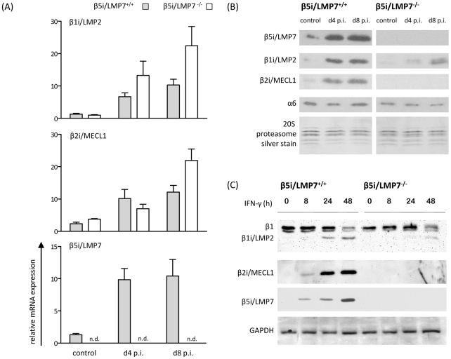
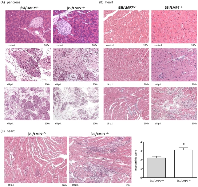
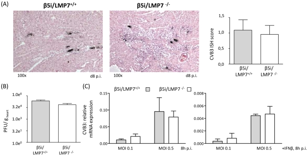
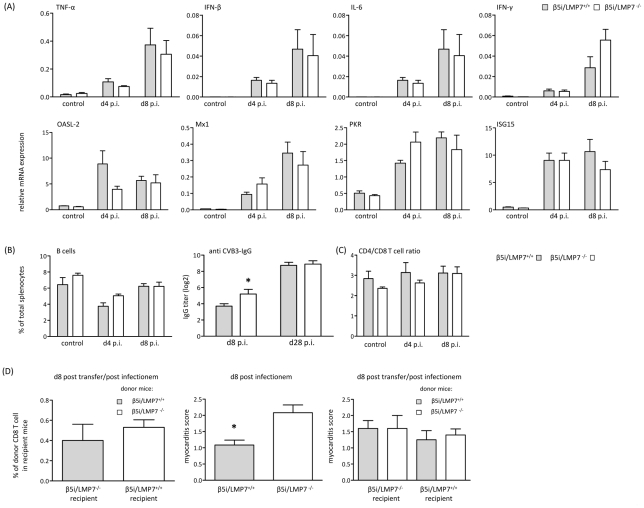
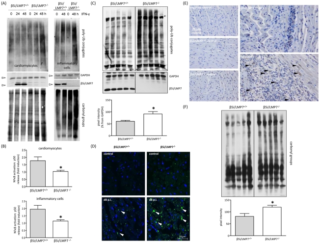
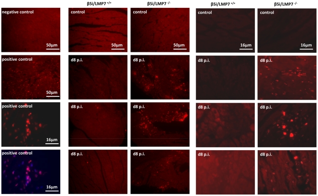
Proteasomes recognize and degrade poly-ubiquitinylated proteins. In infectious disease, cells activated by interferons (IFNs) express three unique catalytic subunits β1i/LMP2, β2i/MECL-1 and β5i/LMP7 forming an alternative proteasome isoform, the immunoproteasome (IP). The in vivo function of IPs in pathogen-induced inflammation is still a matter of controversy. IPs were mainly associated with MHC class I antigen processing. However, recent findings pointed to a more general function of IPs in response to cytokine stress. Here, we report on the role of IPs in acute coxsackievirus B3 (CVB3) myocarditis reflecting one of the most common viral disease entities among young people. Despite identical viral load in both control and IP-deficient mice, IP-deficiency was associated with severe acute heart muscle injury reflected by large foci of inflammatory lesions and severe myocardial tissue damage. Exacerbation of acute heart muscle injury in this host was ascribed to disequilibrium in protein homeostasis in viral heart disease as indicated by the detection of increased proteotoxic stress in cytokine-challenged cardiomyocytes and inflammatory cells from IP-deficient mice. In fact, due to IP-dependent removal of poly-ubiquitinylated protein aggregates in the injured myocardium IPs protected CVB3-challenged mice from oxidant-protein damage. Impaired NFκB activation in IP-deficient cardiomyocytes and inflammatory cells and proteotoxic stress in combination with severe inflammation in CVB3-challenged hearts from IP-deficient mice potentiated apoptotic cell death in this host, thus exacerbating acute tissue damage. Adoptive T cell transfer studies in IP-deficient mice are in agreement with data pointing towards an effective CD8 T cell immune. This study therefore demonstrates that IP formation primarily protects the target organ of CVB3 infection from excessive inflammatory tissue damage in a virus-induced proinflammatory cytokine milieu.


Conflict of interest statement

The authors have declared that no competing interests exist.

Figures

Figure 1
Figure 1. Characterization of 20S proteasomes in IP-deficient mice in CVB3-myocarditis.
(A) Quantitative real-time PCR: β1i/LMP2, β2i/MECL-1, and β5i/LMP7 mRNA expression in the hearts of β5i/LMP7+/+ and β5i/LMP7-/- mice at d4 and d8 p.i. Data shown are representative means of n = 5 mice±SEM. (B) 20S proteasomes were isolated from cardiac tissue and subjected to SDS-PAGE followed by immunoblotting (proteasome subunit α6 is a loading control). Silver staining of isolated 20S proteasomes illustrates equal loading. Data shown are representative for two different 20S proteasome purifications (n = 10 mice per experiment). (C) Primary embryonic cardiomyocytes were stimulated with 100 U/ml IFN-γ for indicated time points and the expression of 20S proteasome immunosubunits was determined in whole tissue homogenates. β1i/LMP2 antibody shows a cross-reaction to constitutive β1 subunit. GAPDH illustrates equal protein loading.
Figure 2
Figure 2. Severe acute CVB3-myocarditis in IP-deficient mice.
Representative hematoxylin-eosin (HE)-staining are illustrated from (A) the pancreas and (B) the heart of β5i/LMP7+/+ and β5i/LMP7-/- mice at different time points p.i. revealing severe acute myocarditis in β5i/LMP7-/- mice at d8 p.i. All cardiac and pancreatic tissue sections shown are representative for at least n = 10 mice/group. (C) Tissue sections from a different experiment confirmed severe heart muscle injury in β5i/LMP7-/- mice at d8 p.i. (enlarged image). To quantify myocardial damage comprising cardiac cell necrosis, inflammation, and scarring, a myocarditis score from 0 to 4 was applied (0: no inflammatory infiltrates, 1: small foci of inflammatory cells between myocytes, 2: larger foci of >100 inflammatory cells, 3: ≤10% of cross-section involved, 4: 10 to 30% of a cross-section involved) (* p<0.05; mean from n = 5 mice).
Figure 3
Figure 3. Characterization of myocardial lesions in acute CVB3-myocarditis.
To characterize inflammatory lesions in CVB3-infected mice in more detail, immunohistology for (A) Mac-3+ macrophages, (B) B220+ B lymphocytes and (C) CD3+ T lymphocytes were performed. Representative cardiac tissue sections are shown, immunohistology was performed for at least n = 6 mice/group. No specific signals were observed in naive control mice. (D) Quantification of immunohistochemically positive cells was achieved by counting all positive cells per visual field at a magnification of x200 in transverse heart tissue sections. Arithmetic means from n ≥5 mice/group±SEM are shown. Differentiation of individual signals for Mac-3 was limited due to massive macrophage infiltration/staining; thus quantitative data are not reflecting absolute macrophage numbers. (E) Quantitative real-time PCR: mRNA expression of CD8 for CD8 T cells, CD4 for CD4 T cells and NKP46 for natural killer cells was determined in the hearts of β5i/LMP7+/+ and β5i/LMP7-/- mice at d8 p.i. Data shown are representative means of n = 5 mice±SEM, * p<0.05.
Figure 4
Figure 4. Viral load is not affected in IP-deficient mice.
(A) The amount of genomic CVB3 RNA as detected by in situ hybridization was scored within a range from 0 to 4 for cardiac tissue sections (n = 10 mice/group). Representative strand-specific in situ hybridizations for CVB3 RNA (black dots) in hearts at the acute stage of myocarditis (d8 p.i.) illustrate equal viral load in both hosts. (B) Plaque assays were performed revealing equal amounts of infectious virus particles in CVB3-infected hearts from β5i/LMP7+/+ and β5i/LMP7-/- mice (n.s.). (C) Primary cardiomyocytes from β5i/LMP7+/+ and β5i/LMP7-/- mice were infected with CVB3 MOI 0.1 and 0.5 for 8 h. To mimic in vivo infection, cardiomyocytes were partially cultured with IFN-β for 24 h prior to CVB3 infection. Viral replication was assessed by quantitative real-time PCR. One representative experiment of three independent experiments is shown.
Figure 5
Figure 5. Adaptive immune responses in CVB3-infected IP-deficient mice.
(A) Quantitative real-time PCR: mRNA expression of indicated cytokines (first row) and antiviral molecules of the innate immune response (second row) was determined in the hearts of β5i/LMP7+/+ and β5i/LMP7-/- mice at d4 and d8 p.i. Data shown are mean of n≥5 mice±SEM. (B)+(C) Flow cytometric analysis was completed on splenocytes isolated from β5i/LMP7+/+ and β5i/LMP7-/- mice at d4 and d8 p.i. (n = 6 mice, representative for 3 independent experiments). (B) Cell numbers of B cells (CD19+, B220+) are illustrated as % of each cell population in comparison to the total number of cells from each spleen (left panel). CVB3-specific IgG levels were determined in sera collected at indicated time points, and titers of virus-specific IgG antibodies were determined by a CVB3-specific ELISA (n≥8 mice, right panel). (C) Ratios of CD4+ T cells (CD4+, CD3+) and CD8+ T cells (CD8+, CD3+) are illustrated. (D) For T cell transfer studies splenic CD8 T cells were separated by MACS (Miltenyi Biotec) from 1×10⁷ splenocytes (Fig. S2B). Left panel: To assess T cell survival after adoptive transfer, CD8 T cells were transferred from CVB3-infected β5i/LMP7+/+ (CD45.1) into β5i/LMP7-/- (CD45.2) mice and from β5i/LMP7-/- mice (CD45.2) into β5i/LMP7+/+ mice (CD45.1). Recipient mice were infected with CVB3 and sacrificed at d8 p.i. T cell survival was determined in splenocytes by flow cytometry. Middle panel: Prior to adoptive T cell transfer, β5i/LMP7+/+ and β5i/LMP7-/- mice were infected with CVB3 and myocarditis scores were determined at d8 p.i. (n = 5 mice). Right panel: Then, splenic CD8 T cells were isolated from these CVB3-infected mice at d8 p.i. as described above; 1–2×10⁶ CD8+ T cells from one donor were injected i.v. through the tail vein into one recipient. Recipient mice were then infected with CVB3 i.p. and sacrificed at d8 p.i. Myocardial damage was evaluated in HE staining as described above. Data shown are mean for n = 5 mice and are representative for two independent experiments.
Figure 6
Figure 6. Accumulation of poly-ub protein conjugates in IP-deficient mice.
(A) Primary cardiomyocytes and B-cell depleted splenocytes from β5i/LMP7+/+ and β5i/LMP7-/- mice were incubated with IFN-γ for indicated time points. Lack of IP formation resulted in accumulation of poly-ub substrates and of oxidant-damaged proteins (staining of carbonyl groups) upon IFN-γ exposure in both cell populations (cell purity is depicted in Fig. S1). GAPDH illustrates equal protein loading. Images are representative from at least two independent experiments. (B) NFκB p50 levels were determined in whole cell homogenates from IFN-γ (100 U/ml for 24 h) treated primary cardiomyocytes and B-cell depleted splenocytes upon 30 min TNF-α exposure. Data are shown as fold increase of p50 levels in comparison to untreated control cells (representative mean from triplicates from at least two independent experiments). (C) Immunoblot of CVB3-infected cardiac homogenates from five individual β5i/LMP7+/+ and β5i/LMP7-/- mice (d8 p.i.) stained for poly-ub-conjugates (from the same transfer; representative for n = 10 mice). Densitometry was performed for individual lanes for these five different mice (normalization to GAPDH control) yielding increased levels of poly-ub signals in β5i/LMP7-/- mice (mean±SEM). (D) Formation of poly-ub containing ALIS (white arrowheads) was visualized by immunofluorescence: heart cryosections were stained with FK1 for poly-ub (green) and Hoechst (blue). Here, slides are representative for n = 6 mice from two independent experiments (more data is illustrated in Fig. S3). (E) To localize poly-ub conjugates within the injured myocardium, immunohistology staining of ubiquitin was performed in control and CVB3-infected mice (d8 p.i.). Representative tissue sections are shown. For more information see Fig. S4 to this figure. (F) CVB3-challenged cardiac lysates from from five individual β5i/LMP7+/+ and β5i/LMP7-/- mice (d8 p.i.; representative for n = 9 mice) stained for oxidant-damaged proteins by staining of carbonyl groups. Quantitative evaluation of oxy-staining was performed as described above. All statistical data are mean±SEM; * p<0.05 as determined by Mann-Whitney U test.
Figure 7
Figure 7. IP-deficient hearts are prone to apoptotic cell death.
Apoptosis was assessed in vivo in cardiac tissue sections in naive and CVB3-challenged mice (d8 p.i.) by in situ cell death detection kit, TMR red. Representative cardiac sections from two independent experiments (infection of n = 5 mice / experiment) are shown. Left panels: controls - upper two slides from first experiment, lower two slides with positive controls from second experiment illustrating DNA strand breaks in inflammatory lesions and surrounding cardiomyocytes, which colocalize to nuclei (merge with Hoechst staining). More information is provided in Fig. S5 + 6 to this figure.

Similar articles

Cited by

References

    1. Watanabe Y, Suzuki O, Haruyama T, Akaike T. Interferon-gamma induces reactive oxygen species and endoplasmic reticulum stress at the hepatic apoptosis. J Cell Biochem. 2003;89:244–253. - PubMed
    1. Si XN, McManus BM, Zhang JC, Yuan J, Cheung C. Pyrrolidine dithiocarbamate reduces coxsackievirus B3 replication through inhibition of the ubiquitin-proteasome pathway. J Virol. 2005;79:8014–8023. - PMC - PubMed
    1. Goldberg AL. Protein degradation and protection against misfolded or damaged proteins. Nature. 2003;426:895–899. - PubMed
    1. Herrmann J, Soares SM, Lerman LO, Lerman A. Potential role of the ubiquitin-proteasome system in atherosclerosis. J Am Coll Cardiol. 2008;51:2003–2010. - PubMed
    1. Patterson C, Ike C, Willis PW, Stouffer GA, Willis MS. The bitter end: the ubiquitin-proteasome system and cardiac dysfunction. Circulation. 2007;115:1456–1463. - PubMed

Publication types

MeSH terms