. 2011 Sep;7(9):e1002235.
doi: 10.1371/journal.ppat.1002235. Epub 2011 Sep 1.

The general transcriptional repressor Tup1 is required for dimorphism and virulence in a fungal plant pathogen

Affiliations

The general transcriptional repressor Tup1 is required for dimorphism and virulence in a fungal plant pathogen

Alberto Elías-Villalobos et al. PLoS Pathog. 2011 Sep.

Abstract

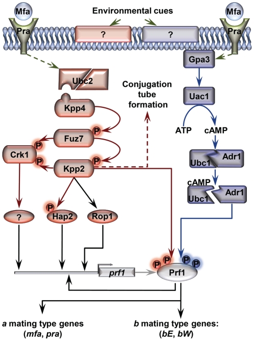
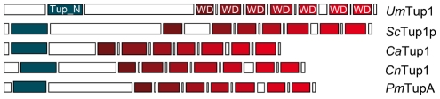
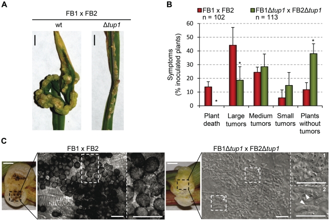
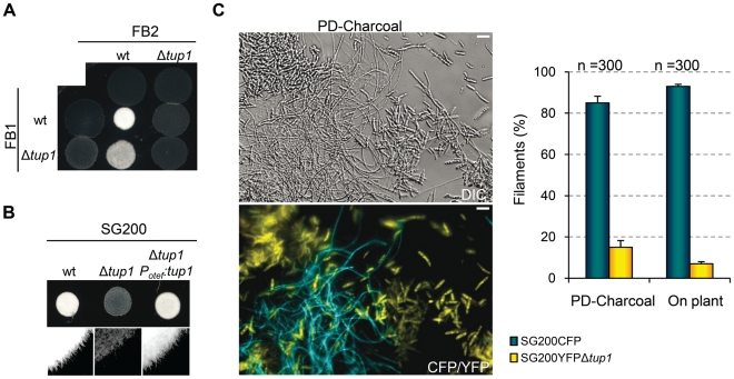
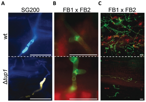
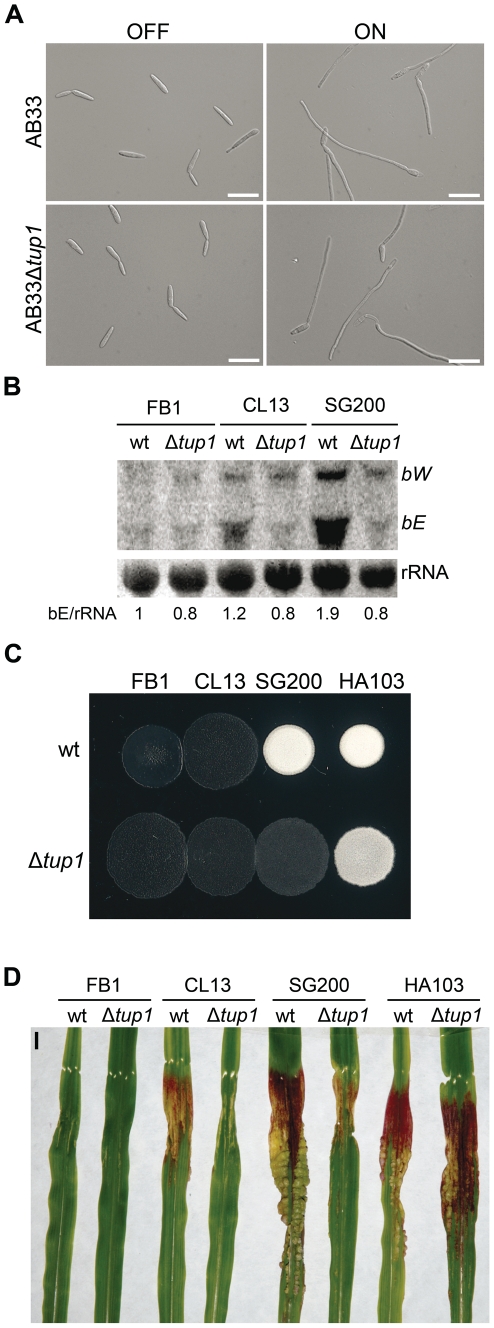
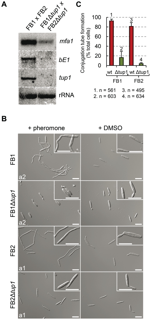
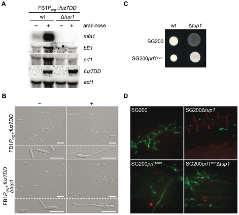
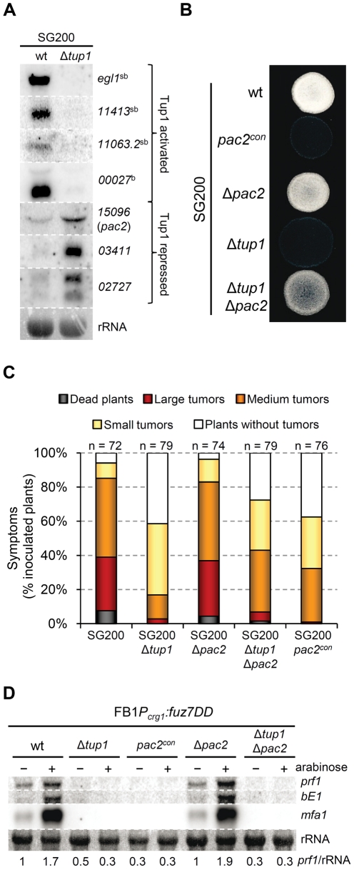
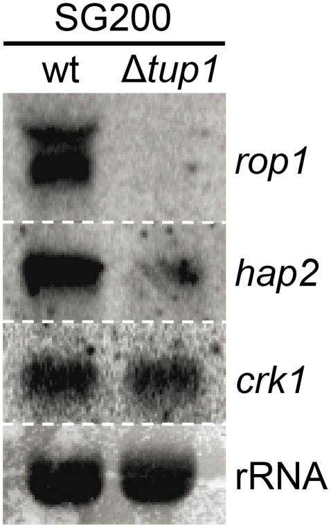
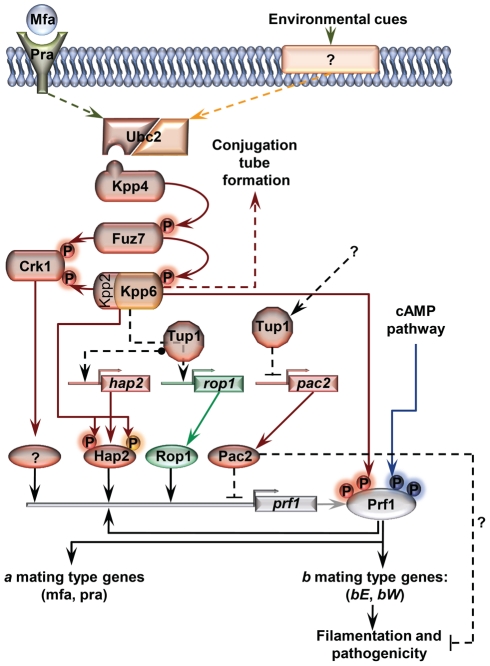
A critical step in the life cycle of many fungal pathogens is the transition between yeast-like growth and the formation of filamentous structures, a process known as dimorphism. This morphological shift, typically triggered by multiple environmental signals, is tightly controlled by complex genetic pathways to ensure successful pathogenic development. In animal pathogenic fungi, one of the best known regulators of dimorphism is the general transcriptional repressor, Tup1. However, the role of Tup1 in fungal dimorphism is completely unknown in plant pathogens. Here we show that Tup1 plays a key role in orchestrating the yeast to hypha transition in the maize pathogen Ustilago maydis. Deletion of the tup1 gene causes a drastic reduction in the mating and filamentation capacity of the fungus, in turn leading to a reduced virulence phenotype. In U. maydis, these processes are controlled by the a and b mating-type loci, whose expression depends on the Prf1 transcription factor. Interestingly, Δtup1 strains show a critical reduction in the expression of prf1 and that of Prf1 target genes at both loci. Moreover, we observed that Tup1 appears to regulate Prf1 activity by controlling the expression of the prf1 transcriptional activators, rop1 and hap2. Additionally, we describe a putative novel prf1 repressor, named Pac2, which seems to be an important target of Tup1 in the control of dimorphism and virulence. Furthermore, we show that Tup1 is required for full pathogenic development since tup1 deletion mutants are unable to complete the sexual cycle. Our findings establish Tup1 as a key factor coordinating dimorphism in the phytopathogen U. maydis and support a conserved role for Tup1 in the control of hypha-specific genes among animal and plant fungal pathogens.


Conflict of interest statement

The authors have declared that no competing interests exist.

Figures

Figure 1
Figure 1. Schematic representation of the regulation of U. maydis mating-type gene expression.
Pheromone (Mfa) recognition by the receptor (Pra) of the opposite mating type, together with environmental cues sensed by unknown receptors (represented by question marks), result in the activation of the cAMP (blue) and MAP kinase (red) pathways. The central core of the MAP kinase module is composed of Kpp4 (MAPKKK), Fuz7 (MAPKK) and Kpp2 (MAPK), and the alternative MAP kinase, Crk1. Once both pathways have been induced, the downstream transcription factor Prf1 becomes transcriptionally and post-translationally activated and the expression of a and b mating-type genes takes place. Transcriptional control of prf1 depends on Rop1, Hap2, a putative unknown factor induced by Crk1, and Prf1 itself. Activation of the MAP kinase module in compatible haploid FB1 or FB2 strains also leads to the formation of conjugation tubes through a Prf1 independent pathway (discontinuous red arrow). Transcriptional regulation is indicated by black arrows. Scheme adapted from .
Figure 2
Figure 2. Comparison of conserved protein domains between different members of the Tup1 family of transcriptional repressors.
Conserved structure of Tup1 proteins in U. maydis (UmTup1), S. cerevisiae (ScTup1p), C. albicans (CaTup1), C. neoformans (CnTup1) and P. marneffei (PmTupA) (for accession numbers see Methods). Domains according to InterPro (Pfam) and functionally characterized in S. cerevisiae , are shown. All the domains described for ScTup1 are conserved in the U. maydis Tup1 protein, including the N-terminal Tup_N domain, required for Ssn6p binding (blue square), seven WD40 domains in the C-terminal region (red tone squares), and a less conserved central region.
Figure 3
Figure 3. tup1 is required for full pathogenic development.
(A) Representative images showing the most prevalent tumor category for wild-type and tup1 mutant infected plants. (B) Disease symptoms caused by wild-type and tup1 mutant strains are shown. Strains are indicated within the color legend. The total number of infected plants (n) is indicated below each strain combination. Symptoms were scored 14 days post-inoculation. Categories correspond to: large tumors (>5 mm), medium tumors (1–5 mm), small tumors (<1 mm). Mean values of three independent experiments and the standard deviation are shown. Asterisk (*) represents statistically significant differences in regard to the wild-type strain. (C) tup1 mutant spore development phenotypes 21 days post-infection. Left: picture of similarly sized tumors developed by the indicated strains. Strong spore formation is evident by dark coloration inside the tumor. Right: tumor sample analyzed by optical microscopy. Spores were present in tumors induced by wild-type strains. Hyphae at fragmentation or rounded cell formation stages were seen (arrowheads) in the tup1 mutant-induced tumors. Mature spores were not observed (scale bar  = 20 µm).
Figure 4
Figure 4. tup1 is required for mating.
(A) Mating between compatible U. maydis strains. The strains indicated (top/left) were spotted either alone or in combination and incubated on PD-charcoal plates for 24 hours at 25°C. A white fuzzy colony appearance is indicative of successful mating and the formation of aerial dikaryotic hyphae. (B) Filament formation in SG200 and SG200Δtup1 strains. The indicated strains were spotted alone on PD-charcoal plates. The presence of white fuzzy colonies indicates the formation of filaments. (C) Quantification of filamentation defects in the tup1 deletion strain. A mixture with equal number of cells from SG200CFP and SG200YFPΔtup1 were spotted onto charcoal plates or inoculated into maize plants. Image on the left represents the filamentation capacity of both strains on charcoal containing media. Scale bar represents 20 µm. The chart on the right indicates the number of filaments that corresponded to each strain in charcoal plates or on the plant leaf surface. Strains are indicated within the color legend. The total number of filaments counted (n) is indicated above each pair of columns. Mean values of three independent experiments and the standard deviation are shown.
Figure 5
Figure 5. Appressorium, clamp-like cells formation and mycelium expansion of tup1 mutants.
(A) Appressorium formed by wild-type SG200CFP and SG200YFPΔtup1 strains. (B) Clamp-like cells formed by wild-type and tup1 mutant cells 2 dpi. (C) Visualization of mycelium expansion inside the plant tissue of the indicated strains 2 dpi. Infected leaf samples were stained with WGA-AF and propidium iodide (see Methods). Scale bars represent 20 µm.
Figure 6
Figure 6. Genetic interaction between tup1 and the b mating-type locus.
(A) Induction of b-compatible heterodimer in the AB33 background. Expression of bE and bW genes was induced by a shift from ammonium (OFF) to nitrate (ON) containing minimal media. b-dependent filament formation could be observed both in wild-type and tup1 mutant strains. Pictures were taken 5 hours post-induction. Scale bars represent 20 µm. (B) b-gene expression level in wild-type and tup1 deletion strains of CL13 (a1 bE1/bW2), SG200 (a1 mfa2 bE1/bW2) and HA103 (a1 (bE1/bW2)con). 10 µg of total RNA extracted from each strain grown on charcoal minimal media for 48 hours at 25°C was loaded per lane. Methylene blue stained rRNA was used as loading control. Numbers indicate the relative signal of bE gene in regard to rRNA. (C) Filamentation capacity of the indicated strains growing on PD-charcoal plates during 24 hours at 25°C. White fuzzy colonies indicates b-dependent filament formation. (D) Representative images showing the most prevalent disease category for wild-type and tup1 mutant infected plants. Strains are indicated below. The FB1 (a1 bE1/bW1) background, which harbors an incompatible b-heterodimer, was used as control. Scale bar represent 1 cm.
Figure 7
Figure 7. Pheromone response and conjugation tube formation in tup1 mutants.
(A) Expression level of mfa1 and bE1 in FB1 vs FB2 and FB1Δtup1 vs FB2Δtup1 crosses after 24 h on charcoal containing plates. tup1 expression was used as experimental control and rRNA used as loading control. (B) DIC images of conjugation hyphae in wild-type and tup1 mutant strains. Wild-type and tup1 mutant strains were grown on CM liquid media until exponential phase and then exposed to the pheromone of the opposite mating type or DMSO (pheromone solvent) for 5 hours. Strains (left), and pheromone or DMSO treatments (top) are indicated. Type of pheromone (a1 or a2) is shown inside each picture. Scale bars indicate 20 µm. (C) Quantification of conjugation hyphae formation in wild-type and tup1 deletion strains. Total number of cells counted is indicated below the chart. Mean values of three independent experiments and the standard deviation are shown.
Figure 8
Figure 8. Tup1-dependent regulation of mating-type genes and prf1 transcription factor.
(A) mfa1, bE1, and prf1 expression levels upon fuz7DD allele induction. Expression of the fuz7DD allele was induced by a shift from a glucose to arabinose containing CM media. Total RNA was extracted 5 hours post-induction and 10 µg were loaded in each lane. U. maydis actin was used as loading control. Strains (above) and probes (right) are indicated. (B) Conjugation tube formation upon fuz7DD induction. Pictures were obtained by optical microscopy 5 hours post-induction. - (glucose) and + (arabinose) indicate non-inducing and inducing conditions, respectively. Scale bars represent 20 µm. (C) Filamentation of constitutive expressed prf1 strains on charcoal media. SG200, SG200prf1con and their derivatives were spotted alone on charcoal plates and grown at 25°C for 24 hours. White fuzzy colonies appearance indicates formation of filaments. (D) On planta filamentation of constitutive expressed prf1 strains. SG200, SG200prf1con and their derivatives were inoculated into maize plants and their filamentation capacity was determined 24 hours post-inoculation.
Figure 9
Figure 9. Microarray validation and pac2 mutants filamentation and virulence phenotypes.
(A) Validation of microarray data by Northern blot. Probes (right) and strains (top) used are indicated. b-dependent (b) and strictly b-dependent genes (sb) according to , are indicated. Methylene blue stained rRNA was used as loading control. Total RNA was extracted from the indicated strains growing on minimal media charcoal-array medium for 48 hours at 25°C. A total of 10 µg of RNA was loaded per lane. (B) Filamentation capacity of the pac2 mutant strains. Strains indicated (left) were spotted alone on PD-charcoal plates and grown for 24 hours at 25°C. pac2 con indicates constitutive expression of pac2 from the otef promoter. (C) Pathogenicity of pac2 mutant strains. Seven day old maize seedlings were infected with the indicated strains (color legend). Total number of infected plants (n) is indicated above each column. Symptoms were scored 14 dpi. Tumors categories correspond to: large tumors (>5 mm), medium tumors (1–5 mm) and small tumors (<1 mm). Represented are the main values of three independent experiments. (D) Pac2-dependent regulation of prf1. Prf1 expression level of the indicated strains upon fuz7DD allele induction was measured by Northern blot. Expression of the fuz7DD allele was induced by a shift from a glucose (-) to arabinose (+) containing CM media. 10 µg of total RNA were loaded per lane. rRNA was used as loading control. Numbers indicate the relative signal of prf1 gene in regard to rRNA.
Figure 10
Figure 10. tup1 is required for wild-type expression levels of the prf1 transcriptional regulators rop1 and hap2.
Northern blot of rop1, hap2 and crk1. 10 µg of total RNA extracted from the indicated strains growing on minimal media charcoal plates for 48 hours at 25°C was loaded per lane. rRNA was used as loading control.
Figure 11
Figure 11. Proposed model for the roles of Tup1 in the control of mating-type genes.
The MAP kinase pathway is shown in red. Black arrows represent transcriptional control. Components exclusively required in laboratory conditions (charcoal, pheromone stimulation, etc) are shown in green. Components specifically required during pathogenesis are shown in orange. In laboratory conditions the effect of Tup1 on prf1 expression would be mediated via its control of hap2, rop1 and pac2 expression levels. During infection, where rop1 is not required, Tup1 would control prf1 expression through hap2 and pac2. Question marks indicate putative elements or interactions.

Similar articles

Cited by

References

    1. Schulz B, Banuett F, Dahl M, Schlesinger R, Schafer W, et al. The b alleles of U. maydis, whose combinations program pathogenic development, code for polypeptides containing a homeodomain-related motif. Cell. 1990;60:295–306. - PubMed
    1. Lo HJ, Kohler JR, DiDomenico B, Loebenberg D, Cacciapuoti A, et al. Nonfilamentous C. albicans mutants are avirulent. Cell. 1997;90:939–949. - PubMed
    1. Lin X, Huang JC, Mitchell TG, Heitman J. Virulence attributes and hyphal growth of C. neoformans are quantitative traits and the MATalpha allele enhances filamentation. PLoS Genet. 2006;2:e187. - PMC - PubMed
    1. Liu H. Co-regulation of pathogenesis with dimorphism and phenotypic switching in candida albicans, a commensal and a pathogen. Int J Med Microbiol. 2002;292:299–311. - PubMed
    1. Lin X. Cryptococcus neoformans: Morphogenesis, infection, and evolution. Infect Genet Evol. 2009;9:401–416. - PubMed

Publication types

MeSH terms